Corpus Callosum Probabilistic Subdivision based on Inter-Hemispheric Connectivity
10 likes | 142 Vues
This study presents a novel automatic probabilistic subdivision model of the Corpus Callosum (CC) utilizing shape analysis and Diffusion Tensor Imaging (DTI) tractography. The model enhances the understanding of regional CC morphology and connectivity by computing subdivision probabilities based on inter-hemispheric fiber characteristics. Results show improved reliability and reproducibility over traditional methods, with significant implications for studying brain development in conditions such as schizophrenia and autism.

Corpus Callosum Probabilistic Subdivision based on Inter-Hemispheric Connectivity
E N D
Presentation Transcript
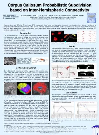
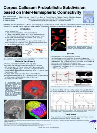
Average Probabilistic Contour Model Corpus Callosum Probabilistic Subdivision based on Inter-Hemispheric Connectivity Martin Styner1,2, Ipek Oguz1, Rachel Gimpel Smith2, Carissa Cascio2, Matthieu Jomier1 1 Department of Computer Science, University of North Carolina at Chapel Hill, 2 Department of Psychiatry, University of North Carolina at Chapel Hill Summary: Use of shape analysis & Diffusion Tensor Imaging (DTI) tractography in order to compute a probabilistic subdivision model of the Corpus Callosum (CC) structure. The CC subdivision allows the study of regional CC morphology for area measurements or DTI properties. Introduction • Corpus callosum (CC): • Major commissural pathway between hemispheres • Integral role in relaying sensory, motor and cognitive information • Much interest in normal development, schizophrenia and autism • Routine subdivision: Witelson[4], motivated by neuro-histology • Bounding Box, AP-length ratios • Often manual: time-consuming, not reproducible and subjective • Novel automatic CC subdivision using probabilistic boundaries based on inter-hemispheric connectivity from DTI[1] 1/2 Fig. 3: Left: Fibers of 4 selected lobes transformed back to MR image space. Middle: Schematic visualization of probability computation. Right: Contour probability maps of 5 training cases. 1/3 1/3 1/5 Results • Probability maps of 5 cases show high similarity • Hard subdivision has low similarity and high decision variability • Largest variability in occipital-temporal section with low probability in a relatively large region. Probabilistic area is relatively large whereas a hard model would highly underestimate this area. • Stability/Reliability (single subject, 10 cases, 5 scanners): ~540mm2 • COV seg: 2.7. COV subdiv: (3.1%,4.1%, 2.4%, 2.5%) • COV manual seg: 5.6% Fig. 1: Corpus callosum in an MR image (left) with Witelson subdivision[5] and its neuro-histological motivation (right). This subdivision scheme is sensitive to alignment and/or manual labeling. Methods/Data/Material • Training population of 5 pediatric cases (age 2-4y) • Automatic lobe subdivision via fluid registration of lobe atlas • CC segmentation: 2D Fourier contour deformable shape model • DTI tractography[2,3] to get a set of inter-hemispheric DTI fibers • Distance-weighted probabilistic subdivision of the CC contour from fiber location in CC • Average of CC subdivision using model correspondence • Probabilities propagated to all CC’s using contour correspondence and Danielsson distance transform based label map • Automatic and stable method Fig. 2: Left below: Midsagittal MR slices and the automatically segmented corpus callosum using a deformable shape model segmentation algorithm. Right below: 3D view of the lobe subdivision (yellow = frontal lobe, purple = parietal lobe, red = occipital lobe, green = temporal lobe). The frontal lobe is further subdivided into an anterior-inferior and a posterior-superior part Right: Set of all inter-hemispheric connectivity from a sample DTI dataset. Only those fibers are considered that connect lobes of the same type. Fig. 4: Left: Final probabilistic subdivision model. Right: Sample subdivision case with relative area noted below. Small study of CC growth in 3 healthy subjects from age 2 to 4 shows CC growth over full length, pronounced in frontal lobe connection region. Fig. 5: Growth curves of CC subdivision regions. Data from 3 healthy subjects from age 2 to age 4. A: Regional growth relative to overall CC growth. B: Regional growth relative to size of corresponding region at age 2. Conclusions Novel automatic, stable probabilistic CC subdivision based on inter-hemispheric connectivity using DTI derived fibers. Original brain images for the corpus callosum were provided by BIOMORPH consortium (EU BIOMED 2) and the UNC Autism Center. This research was supported by the UNC Neurodevelopmental Disorders Research Center HD 03110. REFERENCES [1]. Fillard, P., Gilmore, J., Lin, W., Piven, J., Gerig, G.: Quantitative analysis of white matter fiber properties along geodesic paths. MICCAI. 2879 in Lecture Notes in Computer Science (2003) 16–23 [2]. Xu, D., Mori, S., Solaiyappan, M., van Zijl, P., Davatzikos, C.: A framework for callosal fiber distribution analysis. NeuroImage 17 (2002) 11311143 [3]. Gee, J., Zhang, H., Dubb, A., Avants, B., Yushkevich, P., Duda, J.: Anatomy-based visualizations of diffusion tensor images of brain white matter. Vis. and Image Processing of Tensor Fields. (2005) [4]. Witelson, S.: Hand and sex differences in the isthmus and genu of the human corpus callosum. a postmortem morphological study. Brain 3 (1989) 799–835 [5]. Narr, K., Thompson, P., Sharma, T., Moussai, J., Cannestra, A., Toga, A.: Mapping morphology of the corpus callosum in schizophrenia. Cereb Cortex 1 (2000) 40–9 October 2005, MICCAI 2005 Palm Springs