Download
1 / 1
10 likes | 137 Vues
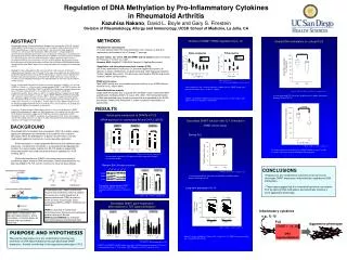
Regulation of DNA Methylation by Pro-Inflammatory Cytokines in Rheumatoid Arthritis Kazuhisa Nakano , David L. Boyle and Gary S. Firestein Division of Rheumatology, Allergy and Immunology, UCSD School of Medicine, La Jolla, CA. n.s. Relative expression. Relative expression.

E N D
Regulation of DNA Methylation by Pro-Inflammatory Cytokines in Rheumatoid Arthritis Kazuhisa Nakano, David L. Boyle and Gary S. Firestein Division of Rheumatology, Allergy and Immunology, UCSD School of Medicine, La Jolla, CA n.s Relative expression Relative expression Relative expression BACKGROUND RESULTS ABSTRACT Global DNA methylation in cultured FLS ** Relative expression Relative expression Rheumatoid arthritis fibroblast-like synoviocytes (RA FLS) exhibit a unique aggressive phenotype that contributes to the cytokine milieu and joint destruction. While the pathogenesis of partial transformation is not fully understood, epigenetic mechanisms are one possible explanation. DNA methylation is a major epigenetic determinant that modulates gene expression, and abnormal methylation is associated with dysregulated cell function. Our recent studies indicate that RA FLS exhibit a unique DNA methylation pattern that could contribute to disease pathogenesis (ACR meeting, 2011) DNA methyltransferases (DNMTs) are critical enzymes involved in establishing proper control of DNA methylation. Global hypomethylation has been described in RA FLS, but the mechanisms have not been defined. * * * * Background/Purpose: Rheumatoid arthritis fibroblast-like synoviocytes (RA FLS) exhibit a unique aggressive phenotype that contributes to the cytokine milieu and joint destruction. While the pathogenesis of partial transformation is not fully understood, epigenetic mechanisms are one possible explanation. DNA methylation is a major epigenetic determinant that modulates gene expression, and abnormal methylation is associated with dysregulated cell function. DNA methyltransferases (DNMTs) are critical enzymes involved in establishing proper control of DNA methylation. Global hypomethylation has been described in RA FLS, but the mechanisms have not been defined. We hypothesized that persistent exposure of pro-inflammatory cytokines can contribute to DNA hypomethylation through decreased of DNMT expression, thereby contributing to the aggressive phenotype of FLS. Method: FLS were obtained from RA and osteoarthritis (OA) synovium at total joint replacement and studied in the 4th through 7th passage. Gene expression was determined by qPCR and protein expression by Western blot analysis. Nuclear extracts and genomic DNA were purified from control or stimulated FLS. DNMT activity was measured with a functional assay and global methylation was determined by an immunoassay that detects methyl-cytosine. Result: Unstimulated RA and OA FLS expressed similar amounts of DNMT1, -3a, and -3b mRNA (n=10 each; n.s.). Western blot showed abundant DNMT1 and DNMT3a protein, but only trace amounts of DNMT3b. DNMT1 and DNMT3a mRNA expression were decreased when FLS were stimulated with IL-1ß (2 ng/ml) for 24 hr (49±8% and 58±6% respectively; p<0.01). Dose responses with IL-1 showed significant suppression of DNMT expression with concentrations as low as 1 pg/ml. DNMT mRNA levels decreased rapidly, with significant suppression after 2 to 8 hrs of IL-1 stimulation. DNMT functional activity was also decreased by IL-1 when FLS were cultured continuously for 14 days with IL-1ß (74.0±6.5% of control, n=11, p=0.0012). 14-days exposure to the DNMT1 inhibitor 5-aza-dC decreased global DNA methylation in OA FLS (78.7±10% of control, n=6, p<0.05), but not in RA FLS (105±6.7% of control, n=6, p=0.22). Conclusion: Exposure to pro-inflammatory cytokines in the synovium decreases DNMT expression and potentially suppresses DNA methylation. The inability of 5-aza-dC to alter RA FLS global methylation suggests that DNMT3a is primarily responsible for DNA methylation in RA, while DNMT1 contributes to methylation OA FLS. These data suggest that the rheumatoid synovium can imprint FLS by altering DNA methylation and potentially inducing a more aggressive phenotype. Basal gene expression of DNMTs in FLS OA RA OA RA mRNA expression in unstimulated RA and OA FLS (qPCR) Med Med Med TNF IL-1 LPS TNF TNF IL-1 LPS IL-1 LPS Poly (I:C) n.s DNMT1 DNMT3a DNMT3b n.s n.s Global DNA methylation (% of standard) Relative expression OA OA RA OA RA RA OA RA Mean ± SD, n=10 each, unpaired t-test Unstimulated RA and OA FLS expressed similar amounts of DNMT1, -3a, and -3b mRNA. Mean ± SD, n=10 RA and n=9 OA lines, un-paired t-test Western Blot (Nuclear extracts) In resting cultured FLS, there was no difference in global methylation between RA and OA FLS. OA 2 OA 3 RA 1 RA 2 RA 3 OA 1 DNMT3a DNMT1 In mammalian cells, DNMTs are the only enzymes that have been shown to mediate the transfer of a methyl group from S-adenosylmethionine (SAM) to cytosine. There are three enzymatically active mammalian DNMTs—DNMT1, DNMT3a and DNMT3b. DNMT1 is primarily a maintenance methyltransferase that preserves methylation patterns during cell division. DNMT3a and DNMT3b can methylate hemimethylated and unmethylated CpG. n.s DNMT1 n.s p=0.036 DNMT3a b-actin Western blot showed abundant DNMT1 and DNMT3a protein. There was no significant difference between RA and OA FLS. Decreased DNMT function after IL-1 stimulation Global DNA methylation (% of standard) When DNA is methylated in the promoter region of genes, where transcription is initiated, genes are silenced and transcription is prevented. (DNMT activity assay) Mean ± SD, , n=6 each, un-paired t-test Poly (I:C) Poly (I:C) Decreased DNMT gene expression after cytokine or TLR ligand stimulation Resting FLS 0 14 DNMT3a DNMT3b PURPOSE AND HYPOTHESIS DNMT1 5-aza-dC (days) n.s Mean ± SD, n = 6 each, paired t-test * Inflammatory cytokines Relative expression We propose that exposure of pro-inflammatory cytokines can contribute to DNA hypomethylation through decreased DNMT expression, thereby contributing to the aggressive phenotype of FLS. * FLS were cultured in the presence of a DNMT inhibitor 5-aza-dC (5 uM). 5-aza-dC significantly decreased global DNA methylation * * * e.g., IL-1b * DNMT activity (OD/h/mg) * * * * FLS IL-1 (ng/ml) Aggressive phenotype DNMT1, 3a, 3b DNA hypomethylation METHODS OA RA Mean ± SD, n = 6 **p < 0.05, *p < 0.01 by ANOVA /Dunnett test Mean ± SD, n=6 each, un-paired t-test Fibroblast-like synoviocytes FLS were obtained from RA and osteoarthritis (OA) synovium at total joint replacement and studied in the 4th through 7th passage. DNMT1 and DNMT3a mRNA expression were significantly decreased when FLS were stimulated with either IL-1ß (2 ng/ml), TNF (50 ng/ml), or LPS (1 ug/ml) for 24 hr. In resting cultured FLS, there was no difference in DNMT function between RA and OA FLS. CONCLUSIONS Nuclear extract (for use in WB and DNMT activity assay) Nuclear extraction kit (Panomics, Fremont, CA, USA) Genomic DNA; MagMAX™ DNA Multi-Sample Kit (Applied Biosystems) Long-term exposure of IL-1b Kinetics of DNMT mRNA regulation by IL-1ß • Exposure to pro-inflammatory cytokines in the synovium decreases DNMT expression and potentially suppresses DNA methylation. • These data suggest that the rheumatoid synovium can imprint FLS by altering DNA methylation and potentially inducing a more aggressive phenotype. Quantitative real-time polymerase chain reaction (PCR) qPCR was performed using Assays On Demand (Applied Biosystems) to determine relative mRNA levels using the GeneAmp 7300 Sequence Detection System (Applied Biosystems). The data were normalized to GAPDH expression to obtain relative cell equivalents. p = 0.008 p = 0.026 Dose-response Time-course DNMT activity (OD/h/mg) * DNMT activity assay Total DNMT activity was assessed by enzymatic activity assay (DNMT Activity/ Inhibition Assay, Active Motif). Relative expression * * Global Methylation analysis Global DNA methylation was evaluated with the MDQ1 Imprint methylated DNA quantification kit (Sigma Aldrich, St. Louis, MO, USA). The methylated fraction of DNA was identified using 5-methylcytosine mAb and quantified by an ELISA-like reaction. Global DNA methylation is shown as percent methylation of a control DNA. * * med IL-1 med IL-1 OA RA Time (hrs) Mean ± SD, n = 4 * p < 0.01 by 2-way ANOVA and contrast testing Mean ± SD, n = 6 each, paired t-test Dose responses with showed significant suppression of DNMT expression with concentrations of IL-1 as low as 1 pg/ml. When FLS were treated for 14 days with 1 ng/ml of IL-1ß, DNMT functional activity was significantly decreased. DNMT1 and DNMT3a mRNA levels began to decrease in 8 and 2 hours after exposure to IL-1ß (2 ng/ml ), respectively.