Download
1 / 59
600 likes | 724 Vues
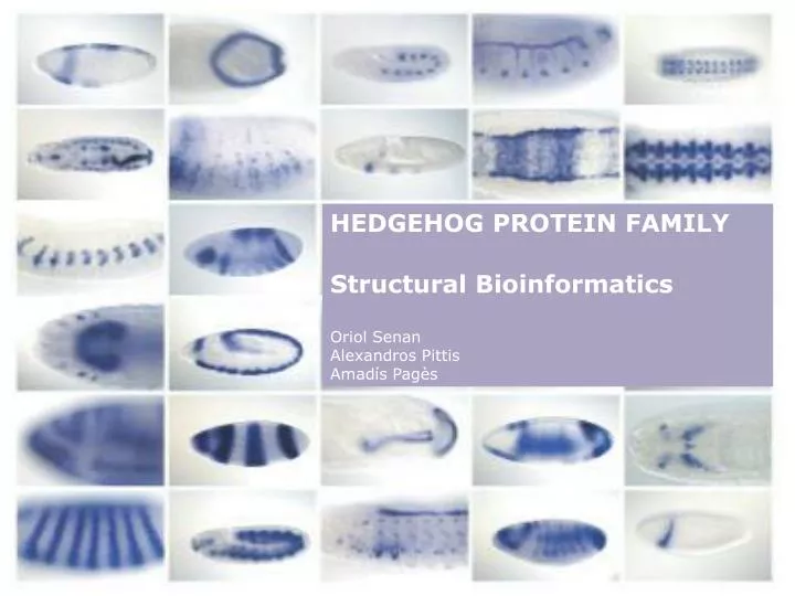
HEDGEHOG PROTEIN FAMILY Structural Bioinformatics Oriol Senan Alexandros Pittis Amadís Pagès. 0. CONTENTS. 1. INTRODUCTION. 2. HEDGEHOG PROTEIN FAMILY. 3. AMINO-TERMINAL HEDGE DOMAIN (HhN). 4. CARBOXY-TERMINAL HOG DOMAIN (HhC). 5. QUESTIONS. 0. CONTENTS. 1. INTRODUCTION.

E N D
HEDGEHOG PROTEIN FAMILY Structural Bioinformatics Oriol Senan Alexandros Pittis Amadís Pagès
0 CONTENTS 1. INTRODUCTION 2. HEDGEHOG PROTEIN FAMILY 3. AMINO-TERMINAL HEDGE DOMAIN (HhN) 4. CARBOXY-TERMINAL HOG DOMAIN (HhC) 5. QUESTIONS
0 CONTENTS 1. INTRODUCTION 2. HEDGEHOG PROTEIN FAMILY 3. AMINO-TERMINAL HEDGE DOMAIN (HhN) 4. CARBOXY-TERMINAL HOG DOMAIN (HhC) 5. QUESTIONS
1 INTRODUCTION • Hedgehog signaling pathway • >Responsible for controled cell growth and differentiation in embryogenesis in metazoa regulating a wide range of patterning events in a local and long-range scale • Left-right asymmetry • Anterior-Posterior patterning • Neural tube patterning • Limb patterning >Homeostatic role in postembryonic tissues (maintenance of stem cell populations) • Continuous Hh pathway activity plays a pathological role in the growth of cancers
1 INTRODUCTION Overview of the Hh signaling pathway > The signal transduction pathway regulate the production of either a transcriptional repressor (CiR) or a transcriptional activator (CiA), active and inactive forms of Ci, a zinc-finger transcription factor > Patched (Ptc) is a 12-transmembrane receptor which acts as an inhibitor for Smoothened (Smo) another transmembrane protein which acts as a signal transducer
1 INTRODUCTION > Downstream of Smo is a multi-protein complex known as the Hedgehog signaling complex (HSC) > Ptc represses Smo, preventing the activation of Hedgehog signalling via proteolytic cleavage of Ci. Cleavage results in a repressor form of Ci, which enters the nucleus and inhibits Hedgehog target gene expression. > The binding of Hedgehog protein to Ptc inactivates it, prevents the inhibition of Smo and induces signal transduction leading to the full length form of Ci (CiA) > The release of the active CiA stimulates the transcription of several target genes
2 THE HEDGEHOG PROTEIN FAMILY 1. INTRODUCTION 2. HEDGEHOG PROTEIN FAMILY 3. AMINO-TERMINAL HEDGE DOMAIN (HhN) 1. > Overview 1. >Structural conformation 1. >Conservation 1. >Interactions with other proteins 4. CARBOXY-TERMINAL HOG DOMAIN (HhC) (oriol) 5. QUESTIONS
2 THE HEDGEHOG PROTEIN FAMILY • > The Hedgehog family consists of secreted signal proteins which comprise different domains • and several motifs • Signal peptide for protein export (SS) • Amino-terminal 'Hedge' secreted signaling domain (HhN) • Carboxy-terminal 'Hog' autocatalytic domain (HnC), containing the 'Hint' module and the Sterol Recognition site (SRR) Signaling domain Autocatalytic domain
2 THE HEDGEHOG PROTEIN FAMILY Role of hedgehog protein in the hedgehog signaling pathway > To become an active ligand requires: > > Autoprocessing reaction > > Palmitoylation of the most amino-terminal cysteine > Once released into the extracellular environment, interacts with different proteins in multimeric form > Targets Patched (Ptc) protein (1) the signal sequence is cleaved Autoprocessing reaction (2) the C-terminal domain of the Hh polypeptide catalyzes an intramolecular cholesteroyl transfer reaction, resulting in (3) the formation of a C-terminally cholesterol-modified Nterminal Hh signaling domain (HhN). This causes association of HhN with membranes, which facilitates the final modification, Palmitoylation (4) the addition of a palmitic acid moiety to the N terminus by an acyltransferase, resulting in the formation of dually modified Hh signaling domain > Skinny hedgehog (Ski or Ras) in Dros. Mel. > Hedgehog AcetylTransferase (HHAT) in mammals
2 THE HEDGEHOG PROTEIN FAMILY Role of the hedgehog protein in the hedgehog signaling pathway > To become an active ligand requires: > > Autoprocessing reaction > > Palmitoylation of the most amino-terminal cysteine > Once released into the extracellular environment, interacts with different proteins in multimeric form > Targets Patched (Ptc) protein Growth Arrest Specific 1 (GAS1) > Attenuates signaling, reduces effective range Hedgehog-Interacting Protein (Hip) > Membrane-bound glycoprotein > Binds to SHH, DHH and IHH > Attenuates signaling, reduces effective range Glypical Dally-Like (Dlp) > Acts as an accessory receptor > Helps in HhN transport Interference Hedgehog (Ihog) - Dros. Mel. Boc / Cdo - Mammals > Required for normal HhN signaling > Facilitate binding to responding cells > Increase HhN association with Ptc
2 THE HEDGEHOG PROTEIN FAMILY Role of the hedgehog protein in the hedgehog signaling pathway > To become an active ligand requires: > > Autoprocessing reaction > > Palmitoylation of the most amino-terminal cysteine > Once released into the extracellular environment, interacts with different proteins in multimeric form > Targets Patched (Ptc) protein The key function of the HhN as an extracellular signal is to inhibit the activity of the receptor Patched (Ptc). In the absence of HhN binding, Patched represses a signaling pathway that acts trough Smoothened. The downstream signaling pathway is ultimately leading Gli (mammals) or Ci (dros. mel.) transcription factors to activate target genes.
2 THE HEDGEHOG PROTEIN FAMILY Dhh • > The Hh gene family is present throughout Eumetazoa although it is absent in some nematodes • One single gene in Drosophila melanogaster • Three paralogous genes in most eumetazoa : • Sonic Hedgehog (Shh), Indian Hedgehog (Ihh) and Desert Hedgehog (Dhh) • Due to genome duplication of ray-finned fishes in zebrafish five hh genes are present >Caenorhabditis elegans has no hh genes but has hh-relates genes via the Hog domain Ihh Shh Drosophila single hh C.elegans hh-like
2 THE HEDGEHOG PROTEIN FAMILY Sequence alignment of Hedgehog family Hedge domain Hog domain
2 THE HEDGEHOG PROTEIN FAMILY • Hedgehog amino-terminal signalling domain 'Hedge' • > The N-terminal domain of Hedgehog proteins • > It has been found in sponges and cnidaria in a large extracellular membrane protein called Hedling • Contains many additional domains apart from Hedge but lacks Hog domain
2 THE HEDGEHOG PROTEIN FAMILY Hedgehog carboxy-terminal autoproteolytic domain 'Hog' >The domain characterizes a group of cysteine peptidases >High similarity of Hint module to self splicing inteins >Several classes of Hint containing proteins, with various types of processing activities
2 AMINO-TERMINAL HEDGE DOMAIN (HhN) Evolution of hh and hh-related genes and domain architecture
3 AMINO-TERMINAL HEDGE DOMAIN (HhN) 1. INTRODUCTION 2. HEDGEHOG PROTEIN FAMILY 3. AMINO-TERMINAL HEDGE DOMAIN (HhN) 1. > Overview 1. >Structural conformation 1. >Conservation 1. >Interactions with other proteins 4. CARBOXY-TERMINAL HOG DOMAIN (HhC) 5. QUESTIONS
HhN -> Overview Domain organization and evolution 3.1 Domain organization and evolution > Present in Hedhegoh proteins > Present in Hedgling proteins > Large extracellular protein that contains a hedge domain at its amino terminus plus many additional > domains such as VWA domains and numerous cadherin repeats, but lacks a Hog domain. > Found in sponges and Cnidaria > Not present in Warthog, Groundhog and Quahog > Proteins which contain a hog domain at its carboxyl terminus but have an amino terminus distinct from HhN. > Found in some nematodes (Caenorhabditis elegans) Mainly hedgehog fragments and very few uncharacterized proteins Hedgehog proteins Hedgling proteins
3.1 HhN -> Overview Domain organization and evolution Domain organization and evolution > Present in Hedhegoh proteins > Present in Hedgling proteins > Large extracellular protein that contains a hedge domain at its amino terminus plus many additional > domains such as VWA domains and numerous cadherin repeats, but lacks a Hog domain. > Found in sponges and Cnidaria > Not present in Warthog, Groundhog and Quahog > Proteins which contain a hog domain at its carboxyl terminus but have an amino terminus distinct from HhN. > Found in some nematodes (Caenorhabditis elegans) Scenario 1 Hedge domain evolved from a secreted amino-terminal domain already associated with the Hog domain. Hedgling is then derived from Hh by a ‘split’ of Hedge from Hog Scenario 2 Hedge domain evolved in a extracellular protein such as hedgling. Then Hedge is ‘merged’ with a Hog protein giving rise to Hh
3.2 HhN -> Structural conformation SCOP Classification Structural conformation • SCOP Classification • Lineage: • Root: scop 2. Class: Alpha and beta proteins (a+b) Mainly antiparallel beta sheets (segregated alpha and beta regions) 3. Fold: Hedgehog/DD-peptidase alpha-beta(2)-alpha-beta(2); 2 layers:alpha/beta Superfamilies: 1. Superfamily: Hedgehog/DD-peptidase zinc-binding motif Families: • Muramoyl-pentapeptide carboxypeptidase • MepA-like • VanX-like • Hedgehog, N-terminal signaling domain • VanY-like
3.2 HhN -> Structural conformation Fold Structural conformation • SCOP Classification • Lineage: • Root: scop 2. Class: Alpha and beta proteins (a+b) Mainly antiparallel beta sheets (segregated alpha and beta regions) 3. Fold: Hedgehog/DD-peptidase alpha-beta(2)-alpha-beta(2); 2 layers:alpha/beta Superfamilies: 1. Superfamily: Hedgehog/DD-peptidase zinc-binding motif Families: • Muramoyl-pentapeptide carboxypeptidase • MepA-like • VanX-like • Hedgehog, N-terminal signaling domain • VanY-like Fold > Core α + β sandwich of two α-helices and six- > stranded mixed β -sheet decorated by extensive > loop regions > Small, two-stranded antiparallel β -sheet
3.2 HhN -> Structural conformation Zinc-binding motif Structural conformation • SCOP Classification • Lineage: • Root: scop 2. Class: Alpha and beta proteins (a+b) Mainly antiparallel beta sheets (segregated alpha and beta regions) 3. Fold: Hedgehog/DD-peptidase alpha-beta(2)-alpha-beta(2); 2 layers:alpha/beta Superfamilies: 1. Superfamily: Hedgehog/DD-peptidase zinc-binding motif Families: • Muramoyl-pentapeptide carboxypeptidase • MepA-like • VanX-like • Hedgehog, N-terminal signaling domain • VanY-like Zinc-binding motif > Zinc coordination site > > Histidine 141 in loop > > Aspartic acid 148 in β-sheet 4 > > Histidine 183 in β -sheet 6 > > Water molecule > > Acid glutamic 177 in β –sheet 5
3.2 HhN -> Structural conformation Zinc-binding motif Structural conformation • SCOP Classification • Lineage: • Root: scop 2. Class: Alpha and beta proteins (a+b) Mainly antiparallel beta sheets (segregated alpha and beta regions) 3. Fold: Hedgehog/DD-peptidase alpha-beta(2)-alpha-beta(2); 2 layers:alpha/beta Superfamilies: 1. Superfamily: Hedgehog/DD-peptidase zinc-binding motif Families: • Muramoyl-pentapeptide carboxypeptidase • MepA-like • VanX-like • Hedgehog, N-terminal signaling domain • VanY-like Zinc-binding motif > Zinc coordination site > > Histidine 141 in loop > > Aspartic acid 148 in β-sheet 4 > > Histidine 183 in β -sheet 6 > > Water molecule > > Acid glutamic 177 in β -sheet 5
3.2 HhN -> Structural conformation Zinc-binding motif Structural conformation • SCOP Classification • Lineage: • Root: scop 2. Class: Alpha and beta proteins (a+b) Mainly antiparallel beta sheets (segregated alpha and beta regions) 3. Fold: Hedgehog/DD-peptidase alpha-beta(2)-alpha-beta(2); 2 layers:alpha/beta Superfamilies: 1. Superfamily: Hedgehog/DD-peptidase zinc-binding motif Families: • Muramoyl-pentapeptide carboxypeptidase • MepA-like • VanX-like • Hedgehog, N-terminal signaling domain • VanY-like Zinc-binding motif > Zinc coordination site > > Histidine 141 in loop > > Aspartic acid 148 in β-sheet 4 > > Histidine 183 in β -sheet 6 > > Water molecule > > Acid glutamic 177 in β -sheet 5
3.2 HhN -> Structural conformation Zinc-binding motif Structural conformation • SCOP Classification • Lineage: • Root: scop 2. Class: Alpha and beta proteins (a+b) Mainly antiparallel beta sheets (segregated alpha and beta regions) 3. Fold: Hedgehog/DD-peptidase alpha-beta(2)-alpha-beta(2); 2 layers:alpha/beta Superfamilies: 1. Superfamily: Hedgehog/DD-peptidase zinc-binding motif Families: • Muramoyl-pentapeptide carboxypeptidase • MepA-like • VanX-like • Hedgehog, N-terminal signaling domain • VanY-like Zinc-binding motif > Zinc coordination site > > Histidine 141 in loop > > Aspartic acid 148 in β-sheet 4 > > Histidine 183 in β -sheet 6 > > Water molecule > > Acid glutamic 177 in β -sheet 5
3.2 HhN -> Structural conformation Zinc-binding motif Structural conformation • SCOP Classification • Lineage: • Root: scop 2. Class: Alpha and beta proteins (a+b) Mainly antiparallel beta sheets (segregated alpha and beta regions) 3. Fold: Hedgehog/DD-peptidase alpha-beta(2)-alpha-beta(2); 2 layers:alpha/beta Superfamilies: 1. Superfamily: Hedgehog/DD-peptidase zinc-binding motif Families: • Muramoyl-pentapeptide carboxypeptidase • MepA-like • VanX-like • Hedgehog, N-terminal signaling domain • VanY-like Zinc-binding motif > Zinc coordination site > > Histidine 141 in loop > > Aspartic acid 148 in β-sheet 4 > > Histidine 183 in β -sheet 6 > > Water molecule > > Acid glutamic 177 in β -sheet 5
3.2 HhN -> Structural conformation Zinc-binding motif Structural conformation • SCOP Classification • Lineage: • Root: scop 2. Class: Alpha and beta proteins (a+b) Mainly antiparallel beta sheets (segregated alpha and beta regions) 3. Fold: Hedgehog/DD-peptidase alpha-beta(2)-alpha-beta(2); 2 layers:alpha/beta Superfamilies: 1. Superfamily: Hedgehog/DD-peptidase zinc-binding motif Families: • Muramoyl-pentapeptide carboxypeptidase • MepA-like • VanX-like • Hedgehog, N-terminal signaling domain • VanY-like Zinc-binding motif > Zinc coordination site > > Histidine 141 in loop > > Aspartic acid 148 in β-sheet 4 > > Histidine 183 in β -sheet 6 > > Water molecule > > Acid glutamic 177 in β -sheet 5
3.2 HhN -> Structural conformation Zinc-binding motif Structural conformation • SCOP Classification • Lineage: • Root: scop 2. Class: Alpha and beta proteins (a+b) Mainly antiparallel beta sheets (segregated alpha and beta regions) 3. Fold: Hedgehog/DD-peptidase alpha-beta(2)-alpha-beta(2); 2 layers:alpha/beta Superfamilies: 1. Superfamily: Hedgehog/DD-peptidase zinc-binding motif Families: • Muramoyl-pentapeptide carboxypeptidase • MepA-like • VanX-like • Hedgehog, N-terminal signaling domain • VanY-like HIS141 HIS183 Zn ASP148 H2O GLU177 Zinc-binding motif > Zinc coordination site > > Histidine 141 in loop > > Aspartic acid 148 in β-sheet 4 > > Histidine 183 in β -sheet 6 > > Water molecule > > Acid glutamic 177 in β -sheet 5
3.3 HhN -> Conservation Sequence Alignment Conservation of the HhN domain in the Hedgehog family of proteins Sequence Alignment Domain is highly conserved, especially in the alpha-beta(2)-alpha-beta(2) region Overall percentages: > Identity percentage: 49% > Similarity percentage: 23%
3.3 HhN -> Conservation Sequence Alignment Conservation of the HhN domain in the Hedgehog family of proteins Sequence Alignment Conservation of residues near the zinc binding motif is really high, with zinc-coordinating histidines and aspartic acid absolutely conserved among vertebrates In Drosophila Melanogaster And Drosophila Hydei there’s only one of those coordinating residues: HIS141 > Hh is not expected to bind zinc > Hh achieves activity in a different > fashion than SHh, IHh and DHh
3.3 HhN -> Conservation Structural Alignment Conservation of the HhN domain in the Hedgehog family of proteins Structural Alignment > 1VHH: SHh – Mus musculus > 2IBG:E: Hh – Drosophila Melanogaster > 2WFQ: DHh – Homo Sapiens > 3HO5:H: SHh – Homo Sapiens
3.3 HhN -> Conservation Structural Alignment Conservation of the HhN domain in the Hedgehog family of proteins Structural Alignment > 1VHH: SHh – Mus musculus > 2IBG:E: Hh – Drosophila Melanogaster sequence identity: 62.66 % pdb1vhh 157 residues -> 4_pdb2ibg 142 residues matching Ca: 140 ( 89.17% / 98.59% )rms deviation: 0.584701 min. length: 6 39 49 59 69 79 89 KLTPLAYKQFIPNVAEKTLGASGRYEGKITRNSERFKELTPNYNPDIIFKDEENTGADRL ************************************************ *** ************************************************ *** YPLVLKQTIPNLSEYTNSASGPLEGVIRRDSPKFKDLVPNYNRDILFR-------DRL 100 110 120 130 140 99 109 119 129 139 149 MTQRCKDKLNALAISVMNQWPGVKLRVTEGWDEDGHHS-EESLHYEGRAVDITTSDRDRS ************************************** * ******************* ************************************** * ******************* MSKRCKEKLNVLAYSVMNEWPGIRLLVTESWDEDYHHGQE-SLHYEGRAVTIATSDRDQS 157 167 177 187 197 207 159 169 KYGMLARLAVEAGFDWVYYESKAHIHCSVKAENSVAAK ******************************** ******************************** KYGMLARLAVEAGFDWVSYVSRRHIYCSVKSD 217 227 237 247 Non cristallized residues
3.4 HhN -> Interactions with other proteins Hh -> Igoh/CDO/BOC Interaction of HhN domain with Ihog/CDO/BOC proteins • Overview of Ihog/CDO/BOC proteins • > Homologous genes • > > Ihog : Drosophila Melanogaster • > > CDO/BOC: Mammals • > Cell-surface proteins • > > Multiple immunoglobulin repeats • > > Multiple fibronectin type 3 (FNIII) repeats • SCOP Classification of interacting domains • Lineage: • root: scop • Class : all beta proteins • Fold : Immunoglobulin-like b sandwich Sandwich; 7 strands in 2 sheets; greek key some members of the fold have additional strands 4. Superfamily : Fibronectin type III 5. Family : Fibronectin type III Protein domains: 2. Fibronectin, different Fn3 modules 30. Brother of CDO precursor 42. Hedgehog receptor iHog Immunoglobulin repeats Fibronectin type III repeats Cell membrane > Ihog binds Hh in the first FNIII repeat : IhogFn1 domain > CDO bind SHh (and homologous genes) in thethird FNIII > repeat : CDOFn3 domain
3.4 HhN -> Interactions with other proteins Hh -> Igoh/CDO/BOC Interaction of HhN domain with Ihog/CDO proteins • Overview of Ihog/CDO proteins • > Homologous genes • > > Ihog : Drosophila Melanogaster • > > CDO (and also BOC) : Mammals • > Cell-surface proteins • > > Multiple immunoglobulin repeats • > > Multiple fibronectin type 3 (FNIII) repeats • SCOP Classification of interacting domains • Lineage: • root: scop • Class : all beta proteins • Fold : Immunoglobulin-like b sandwich Sandwich; 7 strands in 2 sheets; greek key some members of the fold have additional strands 4. Superfamily : Fibronectin type III 5. Family : Fibronectin type III Protein domains: 2. Fibronectin, different Fn3 modules 30. Brother of CDO precursor 42. Hedgehog receptor iHog FNIII repeats of the Ihog protein in Drosophila Melanogaster (IhogFn1-2) Third FNIII repeat of CDO protein in Homo Sapiens (CDOFn3)
3.4 HhN -> Interactions with other proteins Hh -> Igoh/CDO/BOC Interaction of HhN domain with Ihog/CDO proteins Iteraction of Hedgehog and Ihog in Drosophila Melanogaster > IhogFn1-2 forms a symetric dimer complex in the presence of heparin > Each HhN molecule contacts only one single IhogFn1-2 domain > HhN presents an heparin binding site
3.4 HhN -> Interactions with other proteins Hh -> Igoh/CDO/BOC Interaction of HhN domain with Ihog/CDO proteins Iteraction of Hedgehog and Ihog in Drosophila Melanogaster > IhogFn1-2 froms a symetric dimer complex in the presence of heparin > Each HhN molecule contacts only one single IhogFn1-2 domain > HhN presents an heparin binding site V103 V553 L551 Core of three hydrophobic residues surrounded by predominantly polar interactions
3.4 HhN -> Interactions with other proteins Hh -> Igoh/CDO/BOC Interaction of HhN domain with Ihog/CDO proteins Iteraction of Hedgehog and Ihog in Drosophila Melanogaster > IhogFn1-2 froms a symetric dimer complex in the presence of heparin > Each HhN molecule contacts only one single IhogFn1-2 domain > HhN presents an heparin binding site R238 D558 N559 E561 Hydrogen bonds surrounding the hydrophobic core, one through a water molecule
3.4 HhN -> Interactions with other proteins Hh -> Igoh/CDO/BOC Interaction of HhN domain with Ihog/CDO proteins Iteraction of Hedgehog and Ihog in Drosophila Melanogaster > IhogFn1-2 froms a symetric dimer complex in the presence of heparin > Each HhN molecule contacts only one single IhogFn1-2 domain > HhN presents an heparin binding site Key residues in the heparin binding site are highly conserved among invertebrate homologs, but not among vertebrate homologs Vertebrate homologs do not bind via heparin binding site
3.4 HhN -> Interactions with other proteins Hh -> Igoh/CDO/BOC Interaction of HhN domain with Ihog/CDO proteins Iteraction of Sonic Hedgehog and CDO in Homo Sapiens > SHhN presents a calcium binding site. SHhN binds to CDOFn3 in the presence of calcium. > ShhN-CDOFn3 interaction is completely different than Hh-IgohFn1-2 interaction !! Asp96 Asp130 and Asp132 Glu90 and Glu91 Glu127 > Calcium ions are coordinated by three aspartate and three glutamate residues > Each ion is coordinated by eight oxygen atoms
3.4 HhN -> Interactions with other proteins Hh -> Igoh/CDO/BOC Interaction of HhN domain with Ihog/CDO proteins Iteraction of Sonic Hedgehog and CDO in Homo Sapiens > SHhN presents a calcium binding site. SHhN binds to CDOFn3 in the presence of calcium. > ShhN-CDOFn3 interaction is completely different than Hh-IgohFn1-2 interaction !! Glu90 Val198, Met919 and Ile920 His134 > His134 and Glu90 in ShhN make Van der Waals contacts with Val 198, Met919 and Ile920 in CDOFn3 > Mostly acidic residues in CDOFn3 interact with mostly basic residues in SHhN
3.4 HhN -> Interactions with other proteins Hh -> Igoh/CDO/BOC Interaction of HhN domain with Ihog/CDO proteins Iteraction of Sonic Hedgehog and CDO in Homo Sapiens > SHhN presents a calcium binding site. SHhN binds to CDOFn3 in the presence of calcium. > ShhN-CDOFn3 interaction is completely different than Hh-IgohFn1-2 interaction !! > Calcium coordinating residues absolutely > conserved
3.4 HhN -> Interactions with other proteins Hh -> Igoh/CDO/BOC Interaction of HhN domain with Ihog/CDO proteins Iteraction of Sonic Hedgehog and CDO in Homo Sapiens > SHhN presents a calcium binding site. SHhN binds to CDOFn3 in the presence of calcium. > ShhN-CDOFn3 interaction is completely different than Hh-IgohFn1-2 interaction !! > Key residues in the HhN-CDOFn3 interface are > highly conserved > Two non-conserved substitutions: > > Leucine 124 in Drosophila Hydei (expected) > > Leucine 134 in Danio Rerio (unexpected) Conserved substitutions in green
4 CARBOXY-TERMINAL HOG DOMAIN (HhC) 1. INTRODUCTION 2. HEDGEHOG PROTEIN FAMILY 3. AMINO-TERMINAL HEDGE DOMAIN 4. CARBOXY-TERMINAL HOG DOMAIN 1. > Overview 1. >Structural conformation 1. >Structural homologs 1. >Other homologs 5. QUESTIONS
4.1 HhC -> Overview Domain evolution and history HhC components and evolution > Present in Hedhegoh proteins > Present in other proteins families, as a domain homolog. HhC contains the Hint region. Hint superclass. > Distribution in three kingdoms, Bil-A,Bil-B,Bil-C, inteins, hog and Vint. > It seems hog domain has an early origin in eukaryotic evolution. Absence in higher plants. > HhC also contains a SRR region (Sterol recognition region). It binds to cholesterol. This region is also found in other proteins families,but the the nature is unknown. ARR >
4.1 HhC -> Overview Function HhC function > Hint function Mediates de cleavage of the hedge and hog. A aaconserved cysteine mediates a nucleophilic atack on aathe carbonyl of the preceding residue. aaThis includes the formation of a thioester instead of aathe peptide bond. The thioester is atacked by a aacholesterol molecule, and results a aminoterminal aacholesterol modificated residue. > Also contains a SRR region (Sterol recognition aaregion). It binds to cholesterol. This region is also found in other proteins families,but aathe the nature is unknown. Is called ARR(aduct recognition region) >
4.2 HhC -> Structural conformation SCOP Classification Structural conformation • SCOP Classification • Lineage: • Root: scop 2. Class: Alll beta proteins 3. Fold: Hedgehog/intein (Hint) Domain Complex fold made of five beta-hairpin units and a b-ribbon arc Superfamilies: 1. Superfamily: Hedgehog/intein (Hint) domain Families: • Hedgehog, C-terminal (Hog) signaling domain
4.2 HhC -> Structural conformation SCOP Classification Structural conformation (homologues) • SCOP Classification • Lineage: • Root: scop 2. Class: Alll beta proteins 3. Fold: Hedgehog/intein (Hint) Domain Complex fold made of five beta-hairpin units and a b-ribbon arc Superfamilies: 1. Superfamily: Hedgehog/intein (Hint) Domain Duplicated (Contaiins interwined structural repeats Families: • Intein (protein splicing domain)
4.2 HhC -> Structural conformation SCOP Classification Structural conformation (conserved aminoacids) > Several aminoacids crucial for the function
4.2 HhC -> Structural conformation SCOP Classification Structural conformation (conserved aminoacids) > Several aminoacids crucial for the function
4.2 HhC -> Structural conformation Aminoacid conservation Structural conformation (conserved aminoacids) > Cys 258 (in protein Hehgehog Drosophila Does the first nucleophilic atack, crucial for autocleavage