Download
1 / 53
540 likes | 1.06k Vues
Using the Ventilator for More Than Mechanical Ventilation. Joseph E. Previtera, RRT Respiratory Care Department Beth Israel Deaconess Medical Center Boston, MA. The lung in ARDS has three components: Diseased lung that is not recruitable Diseased lung that is recruitable Normal lung.

E N D
Using the Ventilator for More Than Mechanical Ventilation Joseph E. Previtera, RRT Respiratory Care Department Beth Israel Deaconess Medical Center Boston, MA
The lung in ARDS has three components: • Diseased lung that is not recruitable • Diseased lung that is recruitable • Normal lung
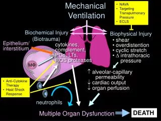
In severe cases of ARDS, no more that 1/3 of all alveoli remain patent. • Large tidal volumes may subject the healthy lung to over-distention and inhibition or inactivation of surfactant • Intense shear forces develop at the junctions of the aerated and non-aerated lung units • The adherent walls of the collapsed small airways often require sustained high pressures to open, but when opened, lower pressures are required to maintain lumenal patency.
The Problem • Atelectasis is bad. • Over inflation is bad. • Alveolar collapse on exhalation is bad. • The ARDS lung is not uniform. • High PEEP is scary. • Auto-PEEP is evil. VILI
Obtaining the P-V Curve • Pre-oxygenate • Record vent settings & signs • Review alarm settings (Apnea setting) • Set resp. rate to 5 bpm • Set PEEP to 0 • Adjust peak flow • Change tidal volume (alternate small-large) • Set plateau for 1-2 sec. interval • Record plateau pressure • Return to previous vent settings and wait 1 min.
To Or not to
1200 Static P-V Curve Technique 1000 800 volume (mL) 600 400 200 0 0 10 20 30 40 pressure (cm H O) pressure manometer 2 calibrated syringe 100 % O2 patient filter
The LIP represents the critical opening pressure of a large • number of alveoli • The UIP reflects the loss of elastic properties of the lung due • to overdistention
Lung Vol. Exhalation Inspiration Pres. PEEP B A Rimensberger et al: The open lung during small tidal volume ventilation: Concepts of recruitment and “optimal” PEEP Crit Care Med; 1999; 27: 1946-1952
Issues with PV Curves • Requires sedation and/or paralysis to measure • Difficult to identify “inflection points” • May require esophageal pressure to separate lung from chest wall effects • Mergoni et al, AJRCCM 1997;156:846-854 • Ranieri et al, AJRCCM 1997;156: 1082-1091 • Deflation limb may be more useful than inflation limb • Holzapfel et al, Crit Care Med 1983; 11: 561-597 • Pressure-volume curves of individual lung units not known
R. SCOTT HARRIS, DEAN R. HESS, and JOSÉ G. VENEGASAm. J. Respir. Crit. Care Med., Volume 161, Number 2, February 2000, 432-439 “There was significant interobserver variability in Pflex, with a maximum difference of 11 cm H2O for the same patient (SD = 1.9 cm H2O)
Beneficial Effects of the “Open Lung Approach” with Low Distending Pressures in Acute Respiratory Distress Syndrome Amato et. Al. Am. J. Respir. Crit. Care Med., Volume 152, pp 1835-1846. 1995
The Problem • Atelectasis is bad. • Over inflation is bad. • Alveolar collapse on exhalation is bad. • The ARDS lung is not uniform. • High PEEP is scary. • Auto-PEEP is evil.
How much PEEP is needed to recruit the lung? • How much PEEP is needed to maintain the lung?
General Approach to Open Lung Technique • Recruit the lung by applying a plateau pressure that can inflate the lung to TLC. • Provide the recruitment pressure for an adequate period of time. • Maintain the lung by not allowing the lung to derecruit on exhalation. • Coming down to the maintenance PEEP level achieves higher lung volumes than titrating up to the maintenance PEEP level.
Patient SelectionPulmonary vs. Extra-Pulmonary ARDS:Gattinoni, Am J Respir Crit Care Med 1998;158:3-11 • Pulmonary ARDS (ARDSP) • Largely consolidation • Little atelectasis • i.e. pneumonia, aspiration, diffuse pulmonary infection, near-drowning, toxic inhalation, lung contusion, etc • Extra-pulmonary ARDS (ARDSEX) • Predominately atelectasis • i.e. sepsis, nonthoracic trauma, pancreatitis, transfusion related injury, etc.
Types of Recruitment Maneuvers “Conventional” • Apneic TLC maneuvers • Non-apneic TLC maneuvers • Prone positioning • Inverse Ratio Ventilation
Patient Monitoring • During recruitment • SpO2 • BP: MAP 60 mm Hg or < 20 mm Hg • HR: > 60 & < 140; no arrhythmia's • After recruitment • VT • Oxygenation
Apneic Lung Recruitment TechniqueA “Conservative” Approach? • Sedation ? • Pre-oxygenation. • CPAP of 30 cm H2O for 30 - 40 seconds. • Monitor Vt and oxygenation for 15 - 30 min. • If unresponsive, repeat at CPAP of 35 to 40 cm H2O.
Non- apneic Lung RecruitmentTechnique:An “Aggressive” Approach? • PCV of 10 - 20 cm H2O. • RR = 10 b/min.; I:E ratio = 1:1. • 3 second IT. • PEEP 20 - 40 cm H2O. • Apply for 45 sec. to 2 min. • Monitor Vt and oxygenation for 15 - 30 min. • If unresponsive, repeat at higher PEEP.
Before recruitment After recruitment Effect of Recruitment: CT Scan
Approaches to Maintain the Recruited Lung Volume • Adequate PEEP • Prone positioning • Sighs
“Open Lung” Management of ARDS: Ventilator Settings • Pressure control ventilation • Tidal volume 6 mL/kg and Pplat 35 cm H2O • PEEP 10 - 20 cm H2O • Usually 15 cm H2O but sometimes higher • FiO2 0.60 (if possible) • Rate 15 - 25/min (avoid auto-PEEP) • IT 1.5 - 2 s (avoid auto-PEEP) • Permissive hypecapnia • Recruitment maneuvers • repeat after each circuit disconnect and as needed. • Prone position
Management of Maintenance PEEP • Reduce FiO2 to 0.60. • Maintain PEEP at lowest level that achieves adequate oxygenation. • Repeat recruitment maneuver if PEEP reduction compromises oxygenation. • Reset PEEP at previous effective level.
PRONE POSITION in ARDS For Every Thing (Turn…Turn…Turn…)
Dorsal Ventral Dorsal Ventral Mechanism of Prone Positioning
PRONE POSITION in ARDS • Proposed Explanations • Increased FRC • Blood Flow Redistribution • Changes in Diaphragmatic Motion • Improved Secretion Removal Magic
Prone Positioning: Procedure • Appropriate staff to manage patient and “tubes”. • 2 - 3 for Airway, IV’s, chest tubes, etc. • 2- 3 for pt. • Minimize abdominal pressure. • Support hips/chest with pillows or deflate abdominal portion of “air bed”. • Maintain pt in Swimming position (one arm extended over head, head turned to that side) • Alternate head/arm Q2o hrs.. • Sedation generally required.
53yo female 1-2 wk flu like syndrome Went to ER unable to breathe 3/18 Intubated in outside hospital Gram + cocci blood/sputum - Strep A Brought to BIDMC 100%, levo, dopa, supine Immediately returned to proned 7.22 46 56 TCPCV 18/15 * 18 .50 1:1.5 3/29 “Pt tolerated supine position for 3 hours today, O2 sats decreased and pt returned to prone position”. 20/15 * 22 .5 7.37 55 88 Vt 500mls
Pappert, D, et al. Influence of Positioning on Ventilation-Perfusion Relationships in Severe ARDS Chest Nov 1994
Prone PositioningChatte. Am J Respir Crit Care Med 1997;155:473:478
Prone Positioning: How Long? Fridrich et al, Anesth Analg 1996;83:1206-1211
Prone Positioning:Clinical ConsiderationsChatte. Am J Respir Crit Care Med 1997;155:473:478 • Duration of proning may need extending. • Increased attention to skin lesions required. • Dependent edema resolves in supine position. • 2 - 4+ personnel required to turn pt. • Special beds not required: • Avoid stiff support; especially under abdomen. • Sedation is usual but not mandatory. • Optimum mode of ventilation is unclear.
Prone Positioning: RisksChatte. Am J Respir Crit Care Med 1997;155:473:478 2/32 pts were intolerant of position alteration • decreased SpO2 of > 5%. 6/32 pts (19%); 294 prone periods (2%) • 2 instances of apical atelectasis. • 1 catheter removal. • 1 catheter compression. • 1 extubation. • 1 transient SVT episode. • Minor skin injury and edema.
The Use of Sighs to Maintain the Open Lung Pelosi, et al. A,J Respir Crit Care Med 1999; 159:872-880. • 3/hr. at a Vt which produces a Pplat of 45 cm H2O. • May open units with opening pressures > than 35 cm H2O. • May resolve absorption atelectasis in poorly ventilated units.
Effect of Sigh on Lung Recruitment and FunctionPelosi. Am J Respir Crit Care Med 1999;159:872-880.
High PEEP Barotrauma Hypotension Reduced cardiac output Increased pulmonary vascular resistance Impaired RV function Permissive Hypercapnia Pulmonary vasoconstriction Myocardial depression Cerebral vasodilatation risk of hemodialysis need for sedation or paralysis Complications of Open Lung Ventilation