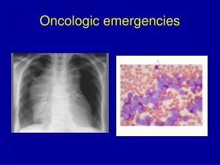
Oncologic challenges in the ED
This Grand Rounds presentation by Dr. Gord McNeil explores the complexities of managing oncologic emergencies in the Emergency Department (ED), focusing on six specific cases encountered in Calgary. Key cases include patients with metastatic spinal cord compression, hemoptysis, and airway obstruction due to cancer. Emphasis is placed on prompt diagnosis, appropriate imaging, and interdisciplinary collaboration for optimal patient outcomes. The discussion highlights evolving treatment protocols and the importance of timely interventions, including surgery and radiation therapy.

Oncologic challenges in the ED
E N D
Presentation Transcript
Oncologic challenges in the ED (besides not getting the old chart from TBCC) Grand Rounds Gord McNeil
6 Cases • Approach • Management • Calgary perspective
Case 1 • 52 year old female with breast cancer presents to the ED with mid back discomfort, progressive weakness of left leg X 1 week and today urinary incontinence • Recent radiation at TBCC (no old chart available)
Approach • Physical • T=37.3 Hr=92, RR=14, BP=172/89 • Decreased sensation left abdominal wall and right lower leg • Decreased power at right knee and ankle • Labs • Hg=109, Plts =302, WBC =6.8, normal lytes and INR.
Differential diagnosis • Epidural abscess • Epidural hematoma • Metastatic spinal cord compression • Routine causes of back pain
Treatment • Dexamethasone IV 10mg prior to MRI, then 4-8 mg q6-8hours • Emergent MRI of entire spine (because pt can have synchronous, multifocal, asymptomatic MSCC.
Treatment • Call Spine service • Decompression of spinal cord is the key to salvage of function • Patchell et al2 in 2005 - radiation for 10 days and decompressive surgery within 24 hours improved outcomes of ambulation, continence and functional abilities from 84% compared to radiation alone for 57%
Metastatic spinal cord compression • Causes • breast(30%), • lung (15%) • prostate (15%) • Other • Sites • thoracic, then lumbar then cervical
MSCC - causes • Expansion of vertebral bone metastasis into epidural space causing cord compression – radiation helps • Neural foramina extension by a paraspinal mass. – radiation helps • Destruction of vertebral cortical bone -requires surgical intervention.
Prognosis • Start of onset of symptoms: Onset: 1-7 days8-14days>14 days Ambulate: 35% 55% 86% (1) • Faster onset = worse prognosis • Start of therapy: dexamethasone and time to surgery • Favorable histology - radiosensitive tumors
Treatment • Radiation only arrests the progression of nonradiosensitive tumors and does not stabilize the spine • Surgery allows immediate cord decompression whereas radiotherapy typically takes several days to weeks.
Calgary perspective • Radiation oncology – Dr. Elizabeth Yan • Radiation did have an important initial role prior to 2005. Now acute surgical decompression and post op radiation is the standard of care.
Calgary perspective • Case scenarios • Highly suspicious for occult CA and back pain then plain films and MRI – no steroids • Known CA and back pain without neuro deficit then MRI, steroids and radiation oncology • Known CA with neuro deficit, then steroids, MRI and spine service
Case 2 • 48 yr old male presents to ED with large hemoptysis X 2 • Recently treated at TBCC for lung CA (old chart not available) • HR =129, RR=32, sat=90% 5L, BP=167/96
Approach • Mobilize team early • Pulmonary • DI/ IR • ICU • Thoracics
Approach • Stabilize • Unstable airway • ETT – large size to faciliate bronchoscope • Not the panacea • Pulmonary toilet – very important • Selective placement of ETT
Approach • Stabilize • CXR –localizes bleeding • Patient position – bleeding side down • Blood products/ fluids prn
Approach • Imaging • CT scan can be done if pt not intubated and has stable airway prior to interventional radiology for bronchial artery emobilization • If ETT then often bronch before IR to localize bleeding
Approach • Hemoglobin not important • patients die of hypoxia not anemia • not like GI bleed
Causes • Friable endobronchial tumors • tumor eroding into a small intrapleural vessel • tumour eroding in to one of the major vessels of the thorax. • Large vessels bleeds = death
Calgary perspective • Dr. Alain Tremblay • One of the few indications for stat call for pulmonary in the middle if the night – involve pulmonary early • Mobilize CT and Interventional radiology early • Supportive management essential
Case 3 • 73 yr old male with thyroid cancer c/o increased secretions, stridor and SOB. • HR = 112, RR=36, BP=178/102, sat=91%on NRB
Approach • Stabilize • O2, suctioning of secretions and allowing patient to sit up • Labs, CXR
Why is it happening? • Usually a subacute process unless an already marginal airway is suddenly compromised by an acute infection, bleeding or the patient’s inability to handle secretions. • Thyroid and esophageal carcinomas may compress the trachea by invading the surrounding soft tissue • Can occur from scarring from prolonged intubation or from radiation therapy
Treatment Consultant • Pulmonary – Rigid scope for endobronchial stenting or laser abalation • Steroids – not helpful (only if known lymphoma)
Calgary perspective • Needs rigid scope • Drs Tremblay and Michaud only 2 pulmonologist in Calgary who do rigid scope (Some thoracic surgeons do as well) • Can call pulmonary at any site and then can help management patient and arrange for rigid scope
Case 4 • 86 yr old female with metastatic lung CA with progressive SOBOE over last 2 weeks, now SOB at rest. • Nonproductive cough, no fever. • HR =92, RR=24, BP 164/92 Sat=94% on 2L
Approach • Stabilize • Labs • CXR • Pleurocentesis
Why is it happening ? • Most common from lung, breast, ovary and lymphoma • Pleural seeding by neoplastic cells increases capillary permeability and produces an exudative effusion • Direct erosion into a blood vessel can cause an abrupt hemorrhagic effusion
Calgary perspective • Dyspnea clinic • Run by Dr. Trembaly and Dr. Michaud • Refer if known CA with symptomatic effusion or if highly suspicious for cancer • Don’t necessarily need tissue diagnosis
Dyspnea Clinic • Tap in ED, send referral. Appt usually in 2 weeks • Clinic places pleurodex catheter and have home care drain it off as necessary • If tapped in ED and return prior to appt, may need admission to pulmonary • Clinic number -521 3511 – Pat Barkley
Case 5 • 64 yr old female with metastatic breast CA to liver “flu –like” symptoms, N/V, lethargy, weakness X 2 weeks • HR =110, RR=16, BP=100/56, Sat =84% RA • GCS = 13, no focal deficit, clinically “dry”
Approach • Labs • Hg =112, WBC =9.4 Plts =186 • Glc = 7.5, Na =132, K = 3.5 • Creatinine =364 (new) • Calcium= 3.64 albumin =29 • Management
Treatment • Measure ionized calcium • ABG • Corrected calcium = measured calcium + (0.02 X(40 – measured albumin) • Lower the albumin and the corrected calcium goes up
Treatment • Replace volume first • Sodium inhibits reabsorption of calcium • Need urine output – 100cc/hr • After euvolemic, then lasix with volume maintenance • Follow K and Mg closely
Causes of hypercalcemia in malignancy • One of the most common complications of cancer - 10-20% • MC caused by breast, lung, renal and cholangiocarcioma and multiple myeloma and lymphoma • Mobilization of bone calcium more rapidly than it can be cleared by the kidneys • Secretion of parathyroid hormone • Presence of bone mets that cause local destruction
Case 6 • 62 yr old male with CML with a recent exacerbation of COPD put on prednisone and levaquin • Acute onset of flank pain then new tonic clonic seizure x 3 minutes • Hr =48, RR =28, BP = 88/52, sat =94%NRB, T=37.6, C/S=6.8
Approach • Stabilize • Labs • Hg = 109, WBC =38, plts=201 • K = 6.8, Na = 132, glc = 6.9 • Cr= 342, urea =32 • Calcium = 1.87, Phosphate = 2.78, albumin =38 • Diagnosis ?
Tumor Lysis Syndrome • Hyperkalemia • Hyperphosphatemia • Hypocalcemia • Renal failure • Renal colic
Tumour lysis syndrome - causes • Large burden of tumor is rapidly and acutely destroyed causes outpouring of potassium, nucleic acids and phosphates. • Sudden build up of electrolytes • MC seen with lymphoma and leukemia, but can also occur with solid organ tumors • Usually within 6 hours to 6 days after the initiation of therapy • Can occur with the administration of corticosteroids to a susceptible patient
Symptoms of hyperkalemia • - weakness and altered MS and arrthymias • Hyperphosphotemia • Causes acute precipitation of calcium in the kidneys and tissues leading to…. • Symptoms of hypocalcemia • carpopedal spasm and seizures • Renal failure • secondary to increased uric acid levels producing renal tubular necrosis • Symptoms of renal colic • secondary to increased uric acid levels producing renal tubular necrosis
Treatment -Tumor lysis syndrome • Aggressive hydration if urine output exists • Alkalinization of urine to pH 7 (can worsen hypocalcemia) • Correct electrolytes and follow closely • Lasix • Allopurinol – 600- 900mg loading dose • Hemodialysis
Rad onc, Med onc, no onc…who goes where? • Radiation therapy • Patient with active radiation – usually gets s/e 2 weeks after starting radiation until 2 weeks after completing radiation – eg diarrhea • Medical oncology • Patient with chemo within the last month • Usually febrile neutropenia at 5 days • No oncology No tissue diagnosis?? – hospitalist