Download
1 / 47
510 likes | 919 Vues
Cell-Cycle Regulation and the Genetics of Cancer. Hartwell Genetics of Cancer. The normal control of cell division Normal cell cycle Molecular signals Machinery Checkpoints that regulate passage through cell cycle How cancer arises from malfunctions in controls over cell division

E N D
Hartwell Genetics of Cancer • The normal control of cell division • Normal cell cycle • Molecular signals • Machinery • Checkpoints that regulate passage through cell cycle • How cancer arises from malfunctions in controls over cell division • Description of cancer phenotypes • Analysis of clonal nature of tumors • Explanation of mutation in protooncogenes and tumor-supressor genes • Comprehensive example describing progression of mutation leading to low-grade brain tumors to aggressive brain cancer
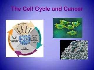
Cancer phenotype results from accumulation of mutations in the clonal progeny of cells • Clone of cells overgrows due to accumulation of mutations controlling proliferation. • Forms tumor and stimulates formation of blood vessels (angiogenesis) • Disseminates through bloodstream to other parts of body (metasteses) • Forms tumors there
General cancer phenotype includes many types of cellular abnormalities • Inappropriate cell-cycle control • Autocrine stimulation – tumor cells make their own signals to divide • Loss of contact inhibition – lost property to stop dividing when contacted by another cell • Loss of cell death – resistance to apoptosis • Loss of gap junctions – no channels for connecting to neighbor cell and communicating directly Feature Figure 18.16 a
Studies based on early inactivation of 1 X chromosome in each cell of women show tumors are each clonal descendents of one cell Fig. 18.18
Cancer arises by successive mutations in a clone of proliferating cells Fig. 18.21
ONCOGENES were first identified in tumor-causing animal viruses: One or 2 viral genes, not the whole virus, was required. The “oncogene” was related to normal cellular genes involved in control of division – often called “protooncogenes” Mutations seen include: Oncogenes (dominant) Tumor suppressor genes (recessive) DNA repair genes Fig. 18.23 b, c
Changes that produce a potential for immortality • Loss of limitations on the number of cell divisions • Ability to grow in culture – normal cells do not grow well in culture on agar plates • Restoration of telomerase activity Feature Figure 18.16 c
Changes that enable tumor to disrupt local tissue and invade distant tissues • Ability to metastasize • Angiogenesis – secrete substances that cause blood vessels to grow toward tumor • Evasion of immune surveillance Feature Figure 18.16 d
Tumor-cell karyotypes often show gross rearrangements Feature Figure 18.16 b (2)
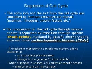
The normal cell division • Cyclin-dependent kinases collaborate with cyclins to ensure the proper timing and sequence of cell-cycle events • The cell cycle has four phases: G1, S G2, and M Fig. 18.2
Cyclin-dependent kinases (CDKs) control the cell cycle by phosphorylating other proteins Fig. 18.7a
Normal gene product must be produced at a particular stage in cell cycle – Yeast mutants: • Cell acquires ability to complete a cell cycle the moment the temperature-sensitive protein has fulfilled its function in that cycle. • CDC28 gene is first step in cell cycle • After CDC28 step, cell is committed to finish cell cycle • Alternative fates after first cell cycle • Arrest of cell division • Fusing with a cell of opposite mating type • Decision to pursue made in G1 phase at “start” Fig. 18.6
CDKs mediate the transition from the G1-to-S phase in human cells Fig. 18.9
Mutations creating defective tumor-suppressor alleles release break on cell division and decrease accuracy of cell reproduction: e.g., retinoblastoma tumor-suppressor gene Fig. 18.24
Target of an enzyme is its substrate • Nuclear lamins • CDK substrates • Underlie inner surface of the nuclear membrane • Probably provide structural support for nucleus • May also be site for assembly of DNA replication, transcription, RNA transport, and chromosome structure proteins • Dissolution of nuclear membrane during mitosis is triggered by CDK phosphorylation of nuclear lamins Fig. 18.7b
Human CDKs and cyclins can function in yeast in place of native proteins Fig. 18.8
Humans make the transition from G2-to-M similar to yeast which is controlled by phosphorylation and dephosphorylation Fig. 18.10
Checkpoints integraterepair of chromosome damage with events of cell cycle • G1-to-S checkpoint • p53 – transcription factor that induces expression of DNA repair genes and CDK inhibitor p21 • p53 pathway activated by ionizing radiation or UV light (causing DNA damage) during G1 phase delays entry into S phase • DNA is repaired before cell cycle continues • If DNA is badly damaged cells commit suicide (programmed cell death or apoptosis) Fig. 18.11 a
Defective Repair: • p53 mutants do not induce p21 and cell cycle is not arrested • Cells replicate damaged DNA • Cells die or DNA is degraded and cell is engulfed and digested by neighboring cells (apoptosis, or programmed cell death) – or: cancer risk increases Fig. 18.11 c,d
Two checkpoints act at the G2-to-M cell-cycle transitiondouble stranded breaks: problems with MITOSIS Fig. 18.12a
Checkpoint in Mspindle damage Fig. 18.12b
Checkpoints ensure genomic stability • Defective checkpoints • Chromosome aberrations • Aneuploidy • Changes in ploidy • Single-stranded nicks – normally repaired in G1 phase • Chromosome loss or gain – normally corrected in G2-to-M checkpoint
Normal cells Cancerous cells Fig. 18.13 b
Three classes of error lead to aneuploidy in tumor cells Fig. 18.13a
Molecular components of each signaling system • Growth factors – hormones and cell-bound signals that stimulate or inhibit cell proliferation • Receptors – membrane bound proteins that accept signals • signal-binding site • transmembrane segment • intracellular domain Fig. 18.15 a
Signal transducers relay messages and transcription factors activate expression of genes Fig. 18.15 b
The protooncogene RAS causes activation of intracellular targets after growth factor binds to receptor – leading to cell replication Fig. 18.15 d
Cancer mutations occur in two forms • Oncogenes • dominant mutations • Mutant tumor-suppressor genes • recessive mutations Fig. 18.22
Oncogenes versus Tumor Suppressor Genes :cancer formation and survival of mice of various genotypes Fig. 18.17
Most cancers result from exposures to mutagens • If one sib or twin gets cancer, other usually does not • Populations that migrate – profile of cancer becomes more like people indigenous to new location
Cancer develops over time Fig. 18.19
Some cancers run in families such as retinoblastoma Fig. 18.20
Two approaches to identifying oncogenes • Analysis of tumor causing retroviruses Fig. 18.23 a
Genetics of brain cancer • Glioblastoma multiforme (GBM) • Aggressive cancer of glial cells • Heterogeneous condition resulting from mutation in different subset of genes • Glial cells • Astrocytes – provide support for neurons • Oligodendrocytes – produce myelin sheaths • Ependymal cells – line the brain cavities known as ventricles and regulate cerebrospinal fluid production • Grades of gliomas • Lowgrade (II) • Anaplastic (III) • GBM (IV) • Low grades progress to higher grades
Many genes in various combinations produce GBMsThree routes for evolution of GBM have been identified Fig. 18.26
Pathway from grade II astrocytoma to malignant GBM Fig. 18.27
Some rapidly arising GBMs have no apparent precursors • Oncogenic amplification of the epidermal-growth-factor-receptor (EGFR) gene and loss of regions from 10p and 10q • Arise de novo or so rapidly no precursors are detectable • Rarely occur in astrocytoma-derived GBM tumors with p53 mutations and 17q deletions • Occur in significantly older adults than GBMs with mutant p53 and chromosome 17 deletions
Summary • GBM phenotypes • Develop by different combinations of mutations in different pathways • Lower-grade astrocytomas via p53 and RB gene inactivations • Oligodendroglial tumors via deletions of chromosome 1 and 19 • de novo via EGFR gene activation • Mutational pathways are often more complicated • Not every GBM shows all genetic changes described • Some GBMs derived from one type of cell have mutations associated with another type of cell
Experiments with yeast helped identify genes that control cell division • Properties of yeast • Grow as haploid or diploid organisms • Can identify recessive mutations in haploids • Complementation analysis in diploids • Budding – daughter cell arises on surface of mother cell and grows in size during cell cycle. Helps determine stage of cell cycle.
Isolation of temperature-sensitive mutants in yeast • Mutants grow normally at permissive temperature • Mutants loses gene function at restrictive temperature • Thousands of cell cycle mutants have been identified Fig. 18.3
A cell-cycle mutant in yeast • (a) growth at permissive temperature displays buds of all sizes • (b) growth at restrictive temperature shows cells have finished first cell cycle and arrested in the second Fig. 18.4
70 cell-cycle genes identified through temperature-sensitive mutation screens
Changes that produce genomic and karyotypic instability • Defects in DNA replication machinery – lost capability to reproduce genome faithfully • Increase rate of chromosomal aberrations – fidelity of chromosome reproduction greatly diminished Feature Figure 18.16 b (1)