Download
1 / 64
650 likes | 868 Vues
BIO 168 NEURAL INTEGRATION II: THE AUTONOMIC NERVOUS SYSTEM AND HIGHER ORDER FUNCTIONS CHAPTER 16. created by Dr. C. Morgan. TOPICS. Introduction and Overview of the ANS The Sympathetic Division The Parasympathetic Division Sympathetic / Parasympathetic Interactions

E N D
BIO 168 NEURAL INTEGRATION II: THE AUTONOMIC NERVOUS SYSTEM AND HIGHER ORDER FUNCTIONS CHAPTER 16 created by Dr. C. Morgan
TOPICS Introduction and Overview of the ANS The Sympathetic Division The Parasympathetic Division Sympathetic / Parasympathetic Interactions Integration and Control Higher Order Functions Brain Chemistry and Behavior Aging and the Nervous System
Introduction and Overview Objectives Review the role of the autonomic nervous system division. Compare the organization of the ANS with the SNS. Examine the structural and functional plan of the ANS.
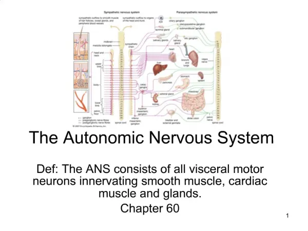
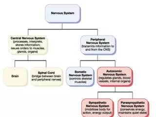
Introduction and Overview If you were unable to think or voluntarily move a muscle, you would continue to survive because your autonomic nervous system (ANS) takes care of vital functions without your conscious awareness or control. Although both the ANS and SNS generate efferent output to effectors, there are important structural differences. In the SNS, lower motor neurons control skeletal muscles. In the ANS, there is a second visceral motor neuron that originates from a synapse in a ganglion outside the CNS. All autonomic nerves are motor although some may also carry visceral sensory information.
Introduction and Overview (cont) SNS plan Upper motor neuron in the cortex. Lower motor neuron in the brain stem or spinal cord. All cell bodies are within the CNS. Fig. 2 a
Introduction and Overview(cont) ANS plan Preganglionic neuron cell body in CNS Postganglionic neuron cell body outside CNS Fig. 2 b
Introduction and Overview(cont) ANS has two subdivisions: sympathetic and parasympathetic, usually with opposing effects if they innervate the same structure or organ. The sympathetic division directs your “fight-or-flight” or crisis responses (protective) with widespread, global output. The parasympathetic division directs your “rest and repose” ongoing visceral responses with localized output. In the sympathetic division, sensory input is consciously perceived but in the parasympathetic division it is not consciously perceived.
Introduction and Overview(cont) The ANS also includes a huge network of neurons and their processes contained within the wall of the digestive tract. This extensive network is sometimes called the enteric nervous system (ENS). This network has as many neurons as the spinal cord and a large variety of neurotransmitters are used. Reflexes within the digestive tract are initiated and controlled locally without commands from the CNS. The ANS does influence the activities along the digestive tract.
TOPICS Introduction and Overview of the ANS The Sympathetic Division The Parasympathetic Division Sympathetic / Parasympathetic Interactions Integration and Control Higher Order Functions Brain Chemistry and Behavior Aging and the Nervous System
Sympathetic Division Objectives Examine the structural arrangement of sympathetic ganglia and their fibers. Using an illustration, explore the distribution of the sympathetic innervation. Discuss the sympathetic chain ganglia and their branching fiber arrangements. Describe the characteristics of sympathetic activation. Discuss neurotransmitter substances and sympathetic function.
Sympathetic Division Sympathetic Division Plan Fig. 3 12
Sympathetic Division (cont) Preganglionic fibers of the sympathetic division are associated with spinal segments T1 to L2. Cell bodies of preganglionic neurons are located in the lateral gray horns of the spinal cord gray matter. Preganglionic neurons synapse with neurons located in ganglia outside the CNS. These ganglia are found in three locations: *in bilateral sympathetic chains adjacent to the spinal column *in peripheral collateral ganglia associated with effectors in the abdominopelvic cavity *in the adrenal medullae tissue proper
Sympathetic Division divergence bilateral Fig. 5
Sympathetic Division (cont) The sympathetic chain includes 3 cervical, 10 – 12 thoracic, 4 – 5 lumbar, 4 – 5 sacral, and 1 coccygeal ganglion. Do not become confused—the cell bodies of preganglionic neurons lie in the spinal cord between T1 and L2 but the sympathetic chain ganglia extend along cervical to sacral segments. There is substantial divergent branching of preganglionic fibers (ascending and descending) so that numerous synapses from neuron collaterals may occur along the chain. It is these fiber branches that produce the chain-like links between the sympathetic (bead-like) ganglia.
Sympathetic Division (cont) Sympathetic ganglia locations via spinal nerves sweat glands, blood vessels, arrector pili muscles, adipose tissue both patterns bilateral (1) Sympathetic chain Fig. 4 a
Sympathetic Division (cont) (2) Passes through chain to collateral ganglion in abdomen Long preganglionic neuron fibers Fig. 4 b
Sympathetic Division (cont) (3) Directly in adrenal medullae Long preganglionic neuron Short postganglionic neuroendocrine cells release epinephrine and norepinephrine Fig. 4 c
Sympathetic Division (cont) Collateral ganglia Celiac: stomach, liver, spleen, pancreas Superior Mesenteric: small intestine Inferior Mesenteric: large intestine, kidneys, bladder, sex organs Postganglionic plexuses Depresses visceral processes
Sympathetic Division (cont) Splanchnic nerves are preganglionic fibers that pass through chain ganglia to synapse in collateral ganglia located in the abdominopelvic cavity. Although splanchnic nerves are bilateral, they serve single ganglia. The ganglia are named for the nearby artery with which they are associated. In the adrenal medulla, preganglionic fibers synapse on neuroendocrine cells which secrete epinephrine and norepinephrine when they are depolarized at the synapse. About 80% of the adrenal output is epinephrine. Sympathetic visceral motor fibers are able to target specific effectors such as blood vessels in the skin.
Sympathetic Division (cont) Sympathetic activation is the response of the entire sympathetic division to a crisis situation. Centers in the hypothalamus direct this response. *RAS is stimulated causing increased alertness, energy, temporary insensitivity to pain, disregard for danger *Pons and medulla direct increased cardiovascular and respiratory responses *Increased muscle tone and perhaps shivering *Mobilization of energy reserves to support increased metabolism Sympathetic activation supports a “fight-or-flight” response.
Sympathetic Division (cont) Postganglionic fibers end in branching telodendria Most varicosities release E and NE but some ganglionic neurons release ACh (sweat glands, smooth muscle of blood vessels in brain and skeletal muscle dilation. of telodendria Fig. 6
Sympathetic Division (cont) 2 types of E and NE neurotransmitter receptors Alpha Alpha 1(1): activates second messenger to excitation Alpha 2 (2 ): reduces cAMP to inhibit cell activity; may inhibit some parasympathetic synapses Beta Beta 1(β1): activates second messenger cAMP to increase cell metabolism ( muscle and liver responses) Beta 2 (β2): opposite of beta 1 so decreases responses action depends on receptors and cellular response TABLE 1
Sympathetic Division (cont) The body wall and skeletal muscle blood vessels are only innervated by the sympathetic division. At most sites, the postganglionic telodendria release ACh which stimulates sweat glands and dilates vasculature in skeletal muscles (muscarinic receptors). In body wall locations, some sympathetic telodendria release NE (1receptor response)which acts to constrict arteries so blood flow is shifted to areas of critical need. Nitric oxide is also released at a few sympathetic synapses involving blood vessel walls of skeletal muscle and the brain. Nitric oxide causes immediate vasodilation.
TOPICS Introduction and Overview of the ANS The Sympathetic Division The Parasympathetic Division Sympathetic / Parasympathetic Interactions Integration and Control Higher Order Functions Brain Chemistry and Behavior Aging and the Nervous System
Parasympathetic Division Objectives Examine the organization of the parasympathetic division. List the general functions of the parasympathetic division. Discuss the parasympathetic neurotransmitter receptors.
Parasympathetic Division Organization little divergence midbrain, pons, medulla lateral gray horns Fig. 7
Parasympathetic Division (cont) CRANIAL PORTION: Preganglionic fibers extend from brain stem nuclei to ganglia. Ganglia associated with cranial nerves III, VII, IX, X Serves lacrimal and salivary glands; intrinsic eye muscles. Vagus (75% of all outflow) to thoracic and abdominal organs (terminal ganglia) SACRAL PORTION: Preganglionic fibers in spinal cord lateral gray horns (S2-S4) with outflow via distinct pelvic nerves (not ventral roots) to intramural ganglia in walls of visceral organs. Innervates kidneys, urinary bladder, terminal large intestine, and sex organs.
Parasympathetic Division (cont) Innervation Plan Preganglionic fibers Ganglia Plexuses Pelvic nerves Vagus nerve*** Fig. 8
Parasympathetic Division (cont) Functions of the parasympathetic division include: *constriction of pupils and nearby focusing of lens *digestive system smooth muscular and secretory activity *promotes hormones secretion supporting anabolic activities *micturition and defecation reflexes *airway constriction *reduction in heart rate and force of contraction *sexual arousal and sex gland stimulation
Parasympathetic Division (cont) ACh and its receptors All parasympathetic synapses use ACh Effects are brief due to AChE and tissue cholinesterase presence. Effects are restricted to specific targets. 2 types of receptors for ACh Nicotinic: Excitatory at all ganglionic synapses Muscarinic:Excitatory or inhibitory at parasympathetic neuromuscular and neuroglandular junctions TABLE 2
TOPICS Introduction and Overview of the ANS The Sympathetic Division The Parasympathetic Division Sympathetic / Parasympathetic Interactions Integration and Control Higher Order Functions Brain Chemistry and Behavior Aging and the Nervous System
Sympathetic / ParasympatheticInteractions Objectives Review the innervation plan of the sympathetic and parasympathetic divisions. Discuss the anatomy of dual innervation. Using an illustration, compare the two divisions. Discuss the concept of autonomic tone.
Sympathetic / Parasympathetic Interactions The sympathetic division is widespread with innervation to nearly all organs and tissues in the body. The parasympathetic division innervates only those structures associated with four cranial nerves and with structures served by the pelvic nerves. Most vital organs receive innervation from both divisions with effects in opposition to one another. This is especially important in the digestive tract, the heart, and the lungs. For the cranial nerves III, VII, IX and X, the postganglionic fibers course along with the cranial nerves while the sympathetic fibers travel from chain ganglia to some of those same structures.
Sympathetic / Parasympathetic Interactions (cont) Sympathetic postganglionic fibers + parasympathetic preganglionic fibers form autonomic plexuses. Fig. 9
Sympathetic / Parasympathetic Interactions (cont) TABLE 2 Comparison Global Local 1:32 1:6 Fig. 10
Sympathetic / Parasympathetic Interactions (cont) In visceral organs where there is dual innervation, there is a certain amount of background output from each division which is called autonomic tone. Some regions and organs are innervated by one division only (blood vessels–sympathetic). Background sympathetic output maintains tone on the peripheral blood vessels so that in order to achieve dilation or constriction, only slight adjustments are needed. TABLE 3 lists a functional comparison of the ANS divisions.
TOPICS Introduction and Overview of the ANS The Sympathetic Division The Parasympathetic Division Sympathetic / Parasympathetic Interactions Integration and Control Higher Order Functions Brain Chemistry and Behavior Aging and the Nervous System
Integration and Control Objectives Introduce the concept of integrating levels of control. Discuss long and short visceral reflexes. Examine higher levels of autonomic control. Discuss the integration of SNS and ANS activities. Describe the concept of biofeedback.
Integration and Control Like the SNS, the ANS is organized into various interacting levels of control. Visceral reflexes represent the lowest level of interaction. Visceral reflexes are comparable to somatic polysynaptic reflexes. Visceral reflexes are either long reflexes or short reflexes. Long reflexes involve the CNS processing of sensory information with ANS motor commands to visceral effectors over a relatively large area or an entire organ. Short reflexes bypass the CNS and synapse on interneurons of autonomic ganglia for localized control of effectors. Short reflexes control activity along small sections of the digestive tract. 40
Integration and Control (cont) Most visceral reflexes are parasympathetic. TABLE 4 41 Fig. 11
Integration and Control (cont) There are centers in the brain stem that control specific functions via the ANS. Simple reflexes cause immediate responses to specific stimuli. Processing centers in the medulla coordinate many complex parasympathetic and sympathetic reflexes. The medulla controls the cardiovascular, respiratory, salivation, swallowing, vomiting, digestive secretory, peristaltic muscular contraction, and urinary functions. The hypothalamus regulates the medullary centers. Because of the widespread interaction of the hypothalamus with other brain nuclei and cortical centers, ANS control and integration becomes very complex.
Integration and Control (cont) Levels of Integration and autonomic control Fig. 12
Integration and Control (cont) SNS and ANS are both under higher system control. Sensory input may result in output via the SNS and ANS simultaneously. TABLE 5 Fig. 13 44
Integration and Control (cont) Biofeedback is an excellent example of higher center control over some ANS functions. Biofeedback is a mechanism that involves monitoring of physiological processes which are “fed back” so a person can invoke conscious thoughts to control them. For example, blood pressure may be monitored. The person is trained to relax and use mechanisms of stress control to lower blood pressure. The monitor informs the subject when pressure drops so they are aware of the success of their control effort. Biofeedback control is used for the management of many disorders.
TOPICS Introduction and Overview of the ANS The Sympathetic Division The Parasympathetic Division Sympathetic / Parasympathetic Interactions Integration and Control Higher Order Functions Brain Chemistry and Behavior Aging and the Nervous System
Higher-Order Functions Objectives Discuss what is meant by higher-order functions. Describe the types of memories. Investigate the brain regions involved in memory consolidation and access. Discuss the cellular mechanisms involved with memory. Describe the state of consciousness. Discuss sleep and arousal from sleep.
Higher-Order Functions Higher-Order functions include: Complex cortical interactions Memory Conscious activities / learning Unconscious activities Because higher-order functions do not involve programmed functions such as reflexes, they are subject to modification and adjustment over time.
Higher-Order Functions (cont) Memories are bits of information stored in the cortex. Fact memories are specific – red stop sign, smell of pizza, shape of a car versus a truck, etc. Skill memories are learned motor behavior patterns – how to open a can, how to turn on the shower, etc. With repetition, skill memories may be become part of your unconscious or programmed behavioral patterns such as eating, driving, swimming, golfing, etc. Cerebral nuclei and the cerebellum will participate.
Higher-Order Functions (cont) Short–term memories are primary, can be recalled immediately, and last a short time. Long–term memories are secondary and last much longer—perhaps even a lifetime. Consolidation converts short term memories to long ones. There are two types of long–term memories: *secondary memories that fade with time; difficult to recall *tertiary memories that are part of your conscious being such as knowing your name, knowing the contours of your body