Download
1 / 26
290 likes | 530 Vues
Fractures and Bone Healing. Dr. Atif Ali Bashir M.D Pathology. Statistics. • Fractures of extremities most common • More common in men up to 45 years of age • More common in women over 45 years of age Before 75 years wrist fractures (Colles’) most common

E N D
Fractures and Bone Healing Dr. Atif Ali Bashir M.D Pathology
Statistics • Fractures of extremities most common • More common in men up to 45 years of age • More common in women over 45 years of age Before 75 years wrist fractures (Colles’) most common • After 75 years hip fractures most common
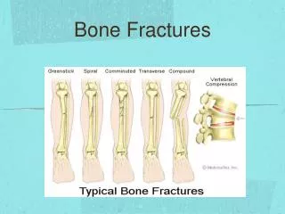
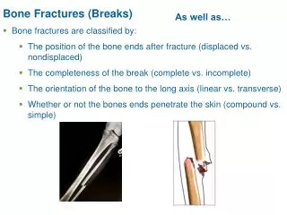
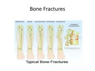
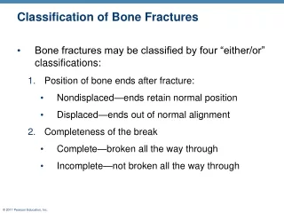
Types of fractures • Magnitude and direction of force • Healthy or diseased bone. • Closed – Bone fragments do not pierce skin • Open/compound – Bone fragments pierce skin • Displaced or undisplaced
Transverse fracture • Usually caused by directly applied force to fracture site
Spiral or Oblique • Caused by violence transmitted through limb from a distance (twisting movements)
Greenstick • Occurs in children: bones soft and bend without fracturing completely
Crush fractures • Fracture in cancellous bone: result of compression (osteoporosis)
Burst fracture • Occurs in short bones, e.g. vertebra from strong direct pressure such as impaction of disc.
Avulsion fracture • Caused by traction, bony fragment usually torn off by a tendon or ligament. • What muscle group attaches to this bony prominence and what nerve also runs in close proximity? • Forearm flexors (common flexor origin) ulnar nerve
Fracture dislocation/subluxation • Fracture involves a joint: results in malalignment of joint surfaces.
Impacted fracture • Bone fragments are impacted into each other.
Comminuated fracture • Two or more bone pieces - high energy trauma
Comminuated fractures can require serious hardware to repair.
Stress fracture • Abnormal stress on normal bone (fatigue fracture) or normal stress on abnormal bone (insufficiency fracture).
C/F of fractures: • Impaired function. • Deformity (unnatural alignment). • Swelling. • Muscle spasm. • Tenderness. • Pain. • Impaired sensation.
Functions of the X-ray • Localises fracture and number of fragments • Indicates degree of displacement • Evidence of pre-existing disease in bone • Foreign bodies or air in tissues • May show other fractures • MRI, CT or ultrasound to reveal soft tissue damage
How to Handle Fractures • Reduction • Open reduction – Allows very accurate reduction – Risk of infection – Usually when internal fixation is needed • Manipulation – Usually with anaesthesia • Traction – Fractures or dislocation requiring slo
Holding the reduction • 4-12 weeks • External fixation • Internal fixation – Intermedually nails, compression plates • Frame fixation
External fixation • Used for fractures that are too unstable for a cast. You can shower and use the hand gently with the external fixator in place.
Frame fixation • Allows correction of deformities by moving the pins in relation to the frame.
Bone Healing 1. Fracture hematoma – blood from broken vessels forms a clot. – 6-8 hours after injury – swelling and inflammation to dead bone cells at fracture site
2. Fibrocartilaginous callus • (lasts about 3 weeks (up to 1st May)) – new capillaries organise fracture hematoma into granulation tissue - ‘procallus’ – Fibroblasts and osteogenic cells invade procallus. – Make collagen fibres which connect ends together – Chondroblasts begin to produce fibrocatilage,
3. Bony callus • (after 3 weeks and lasts about 3-4 months) – osteoblasts make woven bone.
4. Bone Remodeling • Osteoclasts remodel woven bone into compact bone and trabecular bone – Often no trace of fracture line on X-rays.