Protein Sorting
551 likes | 1.65k Vues
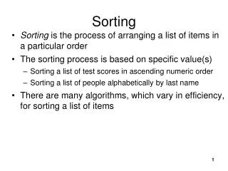
Protein Sorting. Dr. Jason R Mayberry Castle View High School. Functional Organization of a Cell ( Typical Animal Cell). 1 Cytosol. Cytosol. Cytoskeleton providing Structural Support (among other things). Molecules doing the labor for the cell (e.g. ribosomes and other enzymes).

Protein Sorting
E N D
Presentation Transcript
Protein Sorting Dr. Jason R Mayberry Castle View High School
Functional Organization of a Cell (Typical Animal Cell) 1 Cytosol • Cytosol • Cytoskeleton providing Structural Support (among other things) • Molecules doing the labor for the cell (e.g. ribosomes and other enzymes) 2 Endomembrane System • Central Nucleus (an organelle) acting as the information control center Organelles (membrane bound) acting as industrial centers for specific tasks. • Plasma Membrane surrounding the Cell 3 Semiautonomous Organelles • Organelles that have their own DNA, and replicate on their own: mitochondria and chloroplasts
Nucleus Nuclear Pore • 10-20% of cell volume • Nuclear Envelope: Double membrane • Nuclear Pore: allows materials to move in and out (formed where inner and outer membranes come together. • Contains Chromosomes • Chromatin = DNA + histones • Chromosome territory: distinct region occupied by each of the chromosomes. • Nuclear matrix • Nuclear lamina: intermediate filaments linin inner nuclear membrane • Internal matrix: matrix of proteins connected to lamina which organizes the chromosomes • Nucleolus: • dense region inside nucleus where ribosomes are synthesized • rRNA goes directly to nucleolus • Ribosomal proteins are synthesized in cytosol then enter through nuclear pore Chromatin Nuclear Envelope Nucleolus
Nucleus (Figure 4.15) Nucleolus Chromatin Nucleus Pore Nuclear lamina Chromatin Nuclear envelope Pore in nuclear envelope Nucleolus 5.4 µm Pore complexes 0.4 µm Chromatin in nucleus Internal nuclear matrix Inner membrane Nuclear pore complex Nuclear envelope Outer membrane Nuclear lamin proteins Cytosol
Protein Trafficking Protein Trafficking (also called Sorting and Targeting): Process used to get proteins to the proper locations inside the cell, or exported out of the cell • 1) Cytosolic Proteins are produced in the Cytosol and do not need to be sorted. • e.g. Ribosomal Proteins • e.g. Cytoskeletal Proteins • e.g. Proteins for Glycolysis) • 2) Proteins in The following Organelles go through: Post-Translational Sorting • Nucleus • Peroxisome • Mitochondria • Chloroplast • 3) Proteins in the endomembrane system go through: Co-Translational Sorting • Rough ER • Smooth ER • Transport Vesicles • Golgi Apparatus • Vacuoles • Lysosomes • Transmembrane Proteins (in Plasma Membrane and others) • Secretory Vesicles (To be exocytosed)
Where do the Proteins Go? Protein Trafficking (Targeting): getting proteins to the correct location in (or out of) the cell. • 1) Cytosolic Proteins • 1sta.a. don’t form Signaling Sequence • Translation continues in cytosol • Transcription in Nucleus Proteins fold and function within the cytosol • Translation ALWAYS begins in the Cytosol on a freeribosome • 2) Post-translational Sorting: • 1sta.a. form Signal Sequence • Translation completed in cytosol • Polypeptide moved to target organelle by chaperone proteins. chaperone Perixisome Mitochondria Nucleus ER • First several amino acids that are translated determine what happens to the protein during and after translation Chloroplast Hydrophylics.seq. • 3) Co-translational Sorting: • 1sta.a. form Signal Sequence • Translation Pauses as the ribosome moves to and binds ER, and emerging polypeptide fed through pore into ER Lumen. Golgi Hydrophobic s.seq. Additional aa signals determine the final destination with Endomembrane System or out of the Cell.
Endomembrane System and Not Nuclear Envelope double membrane; Nuclear Pores entrance/exit to the nucleus where inner and outer membrane meet. • Mitochondria and Chloroplasts: double membrane; divide and grow on their own; do not fuse with transport vesicles so their membranes are completely independent of the endomembrane system. • Endoplasmic Reticulum • continuous with outer nuclear • Rough ER is studded with ribosomes • Smooth ER is not Transport Vesicles Peroxisomes form by budding off of ER; can also divide in two as they take up materials. Golgi Apparatus Proteins travel in transport vesicles to Cis Face, Medial compartments, Trans Face, then on to other organelles Plasma Membrane: Fuses with transport vesicles from Golgi Lysosome: Forms from transport vesicles from Golgi Vacuole: Fuses with transport vesicles from Golgi (Specializes in different cell types)
Post Translational Sorting A similar mechanism is also used in targeting proteins to Chloroplasts, Peroxisomes, and the Nucleus Note: Mitochondria and Chloroplasts also have their own DNA and produce many of their own proteins; they also produce their own membranes. Cytosol Channel proteins Chaperone Active protein Matrix-targeting sequence Receptor protein Mitochondrial matrix Brooker Fig. 4.31
Co-Translational Sorting SRP: Signal Recognition Peptide Signal peptidase Cytosol 5׳ mRNA NH3+ NH3+ COO– ER signal Sequence Cleaved signal sequence ER membrane SRP receptor Channel protein Additional aa signals determine the final destination with Endomembrane System or out of the Cell. ER lumen From the ER Lumen Transport Vesicle Golgi Transport Vesicle Final Target (Lysosome, Vacuole, Plasma Membrane, or Exocytosis) Brooker: Fig. 4.29
Secretory Pathway Secretory Pathway: Pathway proteins follow from production to exocytosis. 1. Protein enters ER. Ribosome RNA Rough ER 2. Protein exits ER. Protein cis face of Golgi apparatus Golgi apparatus 3. Protein enters Golgi apparatus. 4. Protein exits Golgiapparatus. trans face of Golgi apparatus Plasma membrane 5. Protein is secreted from cell.
Co-translational Sorting of Transmembrane Proteins • Transmembrane proteins go through the Co-translational sorting process • Hydrophobic domains are inserted into the membrane during translation on the Rough ER. SRP: Signal Recognition Peptide Cytosol 5׳ mRNA NH3+
Secretory Pathway Secretory Pathway: Pathway proteins follow from production to exocytosis. 1. Protein enters ER. Ribosome RNA Rough ER 2. Protein exits ER. Protein cis face of Golgi apparatus Golgi apparatus 3. Protein enters Golgi apparatus. 4. Protein exits Golgiapparatus. trans face of Golgi apparatus Transport Protein Molecule being transported Plasma membrane 5. Protein is secreted from cell.
Post Translational Modification of Proteins For proteins to be functional they must often be modified after translation. Cleaving Glycosylation Chaparones • Initiator aa (Methionine) is cleaved off during translation • Signaling aa are cleaved off in ER, Golgi, or target organelle. • Some proteins must be cleaved into smaller pieces to be function (e.g. insulin); Most cleaving takes place in the Golgi • Glycosylation: attachment of a carbohydrate to a Lipid or Protein • Occurs in ERand GolgiApparatus • Most proteins can fold on their own into the ‘right’ (i.e. active) conformation • Chaperones are a class of proteins that help some proteins to fold • Sometimes Chaperones prevent folding until the polypeptide reaches its target organelle (e.g. mitochondria) Chaperone Matrix-targeting sequence
Co-Translational Sorting (Overview) Nucleus Rough ER Smooth ER cis Golgi Plasma membrane trans Golgi
Protein Sorting in the Golgi Apparatus/Complex/Body 1. Proteins bound for different destinations carry distinct tags. Lumen of Golgi apparatus 2. Proteins are sorted. “Tags” 3. Transport vesiclesbud. Receptors Cytosol To other organelle 4. Cytosolic and membraneproteins cause transport vesicles to attach and fuse at destinations. Transport vesicles To plasma membrane for secretion To pre-lysosomal compartment 5. Vesicles deliver contents.
Endomembrane System and Not For any Final Destination… Identify the pathway followed from the beginning of Transcription until a Protein reaches its final destination.
Cotranslational Sorting • Transcription in the Nucleus • Translation begins on Free (Cytosolic) Ribosome • Ribosome|mRNA|PolyPeptide complex moves to Rough ER (Bound Ribosome) where translation completes • Transport Vesicle • Golgi Apparatus (CisFase, medial Comartments, Trans Face) • Transport Vesicle • One of the following • Plasma Membrane • Exocytosis (Extracellular) • Lysosome • Vacuole 1 2 3 4 5 7a 6 7d 7b 7c
Exocytosis and Beyond Moving large items into and out of the cell
Moving LARGER Particles across the Membrane Exocytosis: Movement of materials out of the cell by fusing a vesicle with the plasma membrane SNARE proteins Endocytosis: • Pinocytosis: “Cell Drinking”Brings a small volume of extracellular fluid and the solutes suspended in it into the cell; non Specific • Receptor Mediated EndocytosisReceptors on the cell surface bind specific molecules and are then Endocytosed with the bound molecule. • Phagocytosis: “Cell Eating”Large objects brought into the cell by wrapping the plasma membrane around it then fusing the extensions together.
Endocytosis: Campbell: Figure 7.22 Exocytosis Receptor-Mediated Endocytosis Pinocytosis (Cell drinking) Phagocytosis (Cell eating) EXTRACELLULARFLUID Solutes Pseudopodium Movement of materials out of the cell by fusing a vesicle with the plasma membrane Receptor Ligand Plasmamembrane Coat proteins Coatedpit “Food” orother particle Receptors on the cell surface bind specific molecules and are then Endocytosed with the bound molecule. Proteins lining a portion of the plasma side of the membrane 1) causing it to pinch inward and 2) allowing identification of the contents Coatedvesicle Brings a small volume of extracellular fluid and the solutes suspended in it into the cell; non Specific Vesicle Large objects brought into the cell by wrapping the plasma membrane around it then fusing the extensions together. Foodvacuole
What to do with the stuff brought into the cell • Special organelles are used to break down or detoxify (modify molecularly), materials brought into the cell. • Lysosomes “The Demolition Crew” • Filled with digestive enzymes (hydrolytic and others) • Fuse with endocytic vesicles releasing their enzymes on the contents. • Also used to: • Destroy invading organisms • Break down old and damaged organelles • Facilitate apoptosis (programmed cell death) • Peroxisomes • Filled with enzymes which modify toxic molecules to make them harmless (often via H2O2) • Breakdown of Very Long chain fatty acids • Involved in Myelin formation • Smooth Endoplasmic Reticulum • Continuous with the RER but independent function. • Also detoxifies molecules • Storage (Ca2+) and Production of many molecules • Specialized in many cells to carry out specific functions. • Vacuoles • Central vacuole contribute to Turgor Pressure in plants • Sometimes plays a minor role in cellular “digestion” • Stores water and other materials • Other Functions: Food vacuoles in phagocytes contractile vacuoles in protists, etc.. Smooth Endoplasmic Reticulum Lysosomes Peroxisomes Vacuole Vesicle with large molecules Lysosome Fusion Lysis of molecules
Lysosomes Outside cell Cytosol 1. Receptor-mediated endocytosis uses receptors to bind to macromolecules outside the cell. Plasma membrane pinches in to form a vesicle that delivers cargo to early endosome. Recycling of receptors Receptor-mediated endocytosis 2. Early endosome is acidified and matures into late endosome and, eventually, the lysosome. H+ H+ Early endosome Late endosome Endocytic vesicle 3. Phagocytosis brings smaller cell or food particle inside cell, forming a phagosome. Lysosome Phagocytosis 4.Phagosome is delivered to the lysosome, which fuses with phagosome and digests its contents. 5. Autophagy encloses a damaged organelle within a membrane, forming an autophagosome that is delivered to the lysosome and digested. Phagosome Autophagy 6. Lysosome releases small molecules from digested materials into the cytosol. Autophagosome Damaged organelle
Where Organic Molecules are Made Phospholipids which make up membranes are made on external surface of ER (smooth and rough) CH3 DNA and RNA are made in the nucleus CH3 CH3 CH3(CH2)14COOH CH3 CH3 Proteins are made in the Cytosol and in the Rough ER by Ribosomes either or OH Sugars are made in chloroplasts of photosynthetic organisms Ingested Sugars are broken down in the cytosol and in Mitochondria Ingested Sugars can be stored as Glycogen in the cytosol or Smooth ER Lipids (fats and steroids) are made in the Smooth ER Cytosol
The Cell (Summary) • Plasma Membrane: regulation of materials going in and out of the cell, “sensing” the environment, and cell-cell interactions • Cytosol: basically water contained by the plasma membrane which contains organelles and dissolved molecules. • Organelles • Nucleus: stores DNA and Transcription • Ribosomes: Translation • Rough Endoplasmic Reticulum: Site for translation of proteins to be exported out of the cell (phospholipid synthesis) • Golgi Apparatus: processing and shipping of proteins • Transport vesicles: Transportation of molecules to and from organelles/plasma membrane • Mitochondria: Krebs cycle and Electron Transport chain • Peroxisomes: Detoxification (e.g. H2O2) • Lysosome: “digestion” of large molecules • Vacuole: storage and various functions • Smooth Endoplasmic Reticulum: detoxification, storage, Lipid Synthesis and metabolism • Cytoskeleton • Centrosome: organizing center for Microtubules (contains 2 centrioles) • Cilia: made of microfilaments, used for cell motility and brushing material across the cell surface • Intermediate Filaments: Tensile strength of cells. Smooth Endoplasmic Reticulum Nucleus Intermediate Filaments Ribosomes Rough Endoplasmic Reticulum Lysosomes Golgi Apparatus Centrosome Transport Vesicles Cilia Plasma Membrane Peroxisomes Mitochondria