Download
1 / 53
550 likes | 868 Vues
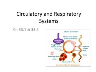
The Circulatory and Respiratory Systems. Chapter 49. Invertebrate Circulatory Systems. Sponges, Cnidarians, and nematodes lack a separate circulatory system - Sponges circulate water using many incurrent pores and one excurrent pore

E N D
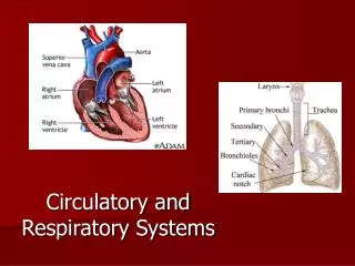
The Circulatory andRespiratory Systems Chapter 49
Invertebrate Circulatory Systems Sponges, Cnidarians, and nematodes lack a separate circulatory system -Sponges circulate water using many incurrent pores and one excurrent pore -Hydra circulates water through a gastrovascular cavity (also for digestion) -Nematodes are thin enough that the digestive tract can also be used as a circulatory system
Invertebrate Circulatory Systems Larger animals require a separate circulatory system for nutrient and waste transport -Open circulatory system = No distinction between circulating and extracellular fluid -Fluid called hemolymph -Closed circulatory system = Distinct circulatory fluid enclosed in blood vessels & transported away from and back to the heart
Vertebrate Circulatory Systems Mammals, birds and crocodilians have a four-chambered heart with two separate atria and two separate ventricles -Right atrium receives deoxygenated blood from the body and delivers it to the right ventricle, which pumps it to the lungs -Left atrium receives oxygenated blood from the lungs and delivers it to the left ventricle, which pumps it to rest of the body
Later-Evolved Vertebrate Circulatory Systems -Oxygenated and deoxygenated blood don’t mix in heart
Heart Valves The heart has two pairs of valves: -Atrioventricular (AV) valves guard the openings between atria and ventricles -Tricuspid valve = On the right -Bicuspid,or mitral, valve = On the left -Semilunar valvesguard the exits from the ventricles to the arterial system -Pulmonary valve = On the right -Aortic valve = On the left
The Cardiac Cycle These valves open and close as the heart goes through the cardiac cycle of rest (diastole) and contraction (systole) -“Lub-dub” sounds heard with stethoscope -Systolic pressure is the peak pressure at which ventricles are contracting AV valves close (to prevent backwash)= “lub” -Diastolic pressure is the minimum pressure between heartbeats at which the ventricles are relaxed, semilunar valves close = (to prevent backwash)“dub” -Blood pressure is written as a ratio of systolic over diastolic pressure
Contraction of Heart Muscle Autorhythmic SA node Initiates depolarization Electrocardiogram (ECG or EKG)
Characteristics of Blood Vessels *Arteries and veins are composed of four tissue layers *Capillaries are composed of only a single layer of endothelial cells...They allow for rapid exchange of gases and metabolites between blood and body cells
Cardiovascular Diseases Atherosclerosis -Accumulation of fatty material within arteries Arteriosclerosis -Arterial hardening due to calcium deposition
Blood Flow and Blood Pressure Blood flow and pressure are regulated by the autonomic nervous system The cardiac center of the medulla oblongata modulates heart rate -Norepinephrine, from sympathetic neurons, increases heart rate -Acetylcholine, from parasympathetic neurons, decreases heart rate
Blood Flow and Blood Pressure Cardiac output is the volume of blood pumped by each ventricle per minute -Increases during exertion because of an increase in both heart rate & stroke volume Arterial blood pressure (BP) depends on the cardiac output (CO) and the resistance (R) to blood flow in the vascular system BP = CO x R
Blood Flow and Blood Pressure The baroreceptor reflex is a negative feedback loop that responds to BP changes -Baroreceptors detect changes in arterial BP -If BP decreases, the number of impulses to cardiac center is decreased -Ultimately resulting in BP increase ...and vice versa.
Veins and Venules Veins and venules -Have thinner layer of smooth muscles than arteries -Return blood to the heart with the help of skeletal muscle contractions and one-way venous valves
What is Blood? A connective tissue composed of a fluid extracellular matrix, called plasma, within which are found different cells and formed elements The functions of circulating blood are: 1. Transportation of materials 2. Regulation of body functions 3. Protection from injury and invasion
The Components of Blood Plasma is 92% water, but it also contains the following solutes: -Nutrients, wastes, and hormones -Ions -Proteins -Albumin,alpha (a) & beta (b) globulins -Fibrinogen -If removed, plasma is called serum
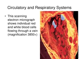
The Components of Blood The formed elements of the blood include red blood cells, white blood cells and platelets Red blood cells (erythrocytes) -About 5 million per microliter of blood -Hematocrit is the fraction of the total blood volume occupied by red blood cells -RBCs of vertebrates contain hemoglobin, a pigment that binds and transports oxygen
White Blood Cells White blood cells (leukocytes) -Less than 1% of blood cells -Larger than erythrocytes and have nuclei -Can also migrate out of capillaries -Granular leukocytes -Neutrophils, eosinophils, and basophils -Agranular leukocytes -Monocytes and lymphocytes
Prothrombin Thrombin Fibrinogen Thrombin Fibrin 3. Cascade of enzymatic reactions is triggered by platelets, plasma factors, and damaged tissue. 1. Vessel is damaged, exposing surrounding tissue to blood. 2. Platelets adhere and become sticky, forming a plug. 5. Once tissue damage is healed, the clot is dissolved. 4. Threads of fibrin trap erythrocytes and form a clot. The Components of Blood Platelets are cell fragments that pinch off from larger cells in the bone marrow -Function in the formation of blood clots
Making Blood “Formed Elements” All of the “formed elements” develop from pluripotent stem cells Hematopoiesisis blood cell production -Occurs in the bone marrow of (medullary bone), and produces: -Lymphoid stem cell...Lymphocytes -Myeloid stem cell...All other blood cells Red blood cell production is called erythropoiesis
Lungs Gills were replaced in terrestrial animals because 1. Air is less supportive than water 2. Water evaporates The lung minimizes evaporation by moving air through a branched tubular passage
Gas Exchange Gases diffuse directly into unicellular organisms However, most multicellular animals require system adaptations to enhance gas exchange -Amphibians respire across their skin -Echinoderms have protruding papulae -Insects have an extensive tracheal system -Fish use gills -Mammals have a large network of alveoli
Gills Gills are specialized extensions of tissue that project into water External gills are not enclosed within body structures -Found in immature fish and amphibians -Two main disadvantages -Must be constantly moved to ensure contact with oxygen-rich fresh water -Are easily damaged
Gills The gills of bony fishes are located between the oral (buccal or mouth) cavity and the opercular cavities -These two sets of cavities function as pumps that alternately expand -Moving water into the mouth, through the gills, and out of the fish through the open operculum or gill cover
Gills There are four gill arches on each side of a fish’s head -Each is composed of two rows of gill filaments, which consist of lamellae -Within each lamella, blood flows opposite to direction of water movement -Countercurrent flow -Maximizes oxygenation of blood
D A Dp R = d Gas Exchange The rate of diffusion between two regions is governed by Fick’s law of diffusion R = Rate of diffusion D = Diffusion constant A = Area over which diffusion takes places Dp = Pressure difference between two sides d = Distance over which diffusion occurs
Lungs -Lungs of mammals are packed with millions of alveoli (sites of gas exchange) -Inhaled air passes through the larynx, glottis and trachea -Bifurcates into the right and left bronchi, which enter each lung and further subdivide into bronchioles -Surrounded by an extensive capillary network
Lungs Air exerts a pressure downward, due to gravity -A pressure of 760 mm Hg is defined as one atmosphere (1.0 atm) of pressure Partial pressure is the pressure contributed by a gas to the total atmospheric pressure -Based on the % of the gas in dry air -PN2 = 760 x 79.02% = 600.6 mm Hg -PO2 = 760 x 20.95% = 159.2 mm Hg -PCO2 = 760 x 0.03% = 0.2 mm Hg
Lungs Lungs of amphibians are formed as saclike outpouchings of the gut Frogs have positive pressure breathing -Force air into their lungs by creating a positive pressure in the buccal cavity Reptiles have negative pressure breathing -Expand rib cages by muscular contractions, creating lower pressure inside the lungs
Lung Structure and Function During inhalation, thoracic volume increases through contraction of two muscle sets -Contraction of the external intercostal muscles expands the rib cage -Contraction of the diaphragm expands the volume of thorax and lungs -Produces negative pressure which draws air into the lungs
Lung Structure and Function Tidal volume = Volume of air moving in and out of lungs in a person at rest Vital capacity = Maximum amount of air that can be expired after a forceful inspiration Hypoventilation = Insufficient breathing -Blood has abnormally high PCO2 Hyperventilation = Excessive breathing -Blood has abnormally low PCO2
Lung Structure and Function Each breath is initiated by neurons in a respiratory control center in the medulla oblongata -Stimulate external intercostal muscles and diaphragm to contract, causing inhalation -When neurons stop producing impulses, respiratory muscles relax, and exhalation occurs
Gas Exchange Gas exchange is driven by differences in partial pressures -As a result of gas exchange in the lungs, systemic arteries carry oxygenated blood with relatively low CO2 concentration -After the oxygen is unloaded to the tissues, systemic veins carry deoxygenated blood with a high CO2 concentration
Lung Structure and Function Neurons are sensitive to blood PCO2changes -A rise in PCO2 causes increased production of carbonic acid (H2CO3), lowering the pH -Stimulates chemosensitive neurons in the aortic and carotid bodies -Send impulses to control center Brain also contains central chemoreceptors that are sensitive to changes in the pH of cerebrospinal fluid (CSF)
Lung Structure and Function -Each breath is initiated by neurons in a respiratory control center in the medulla oblongata
Respiratory Diseases Chronic obstructive pulmonary disease (COPD) refers to any disorder that obstructs airflow on a long-term basis -Asthma = An allergen triggers the release of histamine, causing intense constriction of the bronchi and sometimes suffocation -Emphysema = Alveolar walls break down and the lung exhibits larger but fewer alveoli -Lungs become less elastic
Respiratory Diseases Lung cancer follows or accompanies COPD -The number one cancer killer -Caused mainly by cigarette smoking
RBCs of vertebrates contain Hemoglobin, a pigment that binds and transports oxygen Hemoglobin consists of four polypeptide chains: two a and two b -Each chain is associated with a heme group, and each heme group has a central iron atom that can bind a molecule of O2 Hemoglobin loads up with oxygen in the lungs, forming oxyhemoglobin -Some molecules loseO2as blood passesin capillaries, forming deoxyhemoglobin
Hemoglobin In a person at rest, about one-fifth of the oxygen is unloaded in the tissues -Leaving four-fifths of the oxygen in the blood as a reserve -This reserve enables the blood to supply body’s oxygen needs during exertion The oxyhemoglobin dissociation curve is a graphic representation of these changes
Hemoglobin Hemoglobin’s affinity for O2 is affected by pH and temperature -The pH effect is known as the Bohr shift -Caused by H+ binding to hemoglobin -Results in a shift of oxyhemoglobin dissociation curve to the right -Facilitates oxygen unloading -Increasing temperature has a similar effect
-Mitochondria release CO2 after pyruvate oxidation and the Krebs CycleMitochondria also reduce oxygen to H20 (water) at the end of the electron transport chain. C02 leaves tissue cells Gas Exchange Biochemistry In Tissue Cells