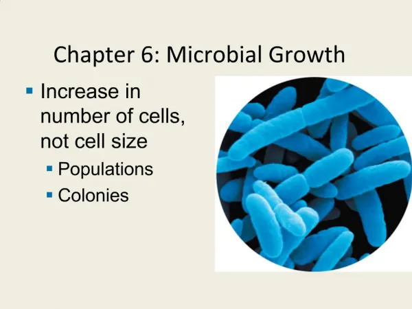
Ch 6 Microbial Growth
Ch 6 Microbial Growth. Student Learning Outcomes: Classify microbes into five groups on the basis of preferred temperature range. Explain the importance of osmotic pressure to microbial growth.

Ch 6 Microbial Growth
E N D
Presentation Transcript
Student Learning Outcomes: Classify microbes into five groups on the basis of preferred temperature range. Explain the importance of osmotic pressure to microbial growth. Provide a use for each of the four elements (C, N, S, P) needed in large amounts for microbial growth. Explain how microbes are classified on the basis of O2 needs. Identify ways in which aerobes avoid damage by toxic forms of O2. Describe the formation of biofilms and their potential for causing infection. Distinguish between chemically defined and complex media. Justify the use of each of the following: anaerobic techniques, living host cells, candle jars, selective, differential, and enrichment media. Define colony and CFUs and describe how pure cultures can be isolated with streak plates. Explain how microbes are preserved by deep-freezing and lyophilization. Distinguish between binary fission and budding. Define generation time and explain the bacterial growth curve. Review some direct and indirect methods of measuring bacterial cell growth.
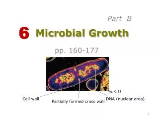
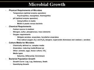
Microbial Growth Microbial growth: Increase in cell number, not cell size! Physical Requirements for Growth: Temperature • Minimum growth temperature • Optimum growth temperature • Maximum growth temperature Five groups based on optimum growth temperature • Psychrophiles • Psychrotrophs • Mesophiles • Thermophiles • Hyperthermophiles Figs. 6 .2 & 3
Figs 6.2/3:Food preservation temperatures andEffect of amount of food on its cooling rate
Physical Requirements for Growth:pH and Osmotic Pressure Most bacteria grow best between pH 6.5 and 7.5: Neutrophils Some bacteria are very tolerant of acidity or thrive in it: Acidophiles(preferred pH range 1 to 5) Molds and yeasts grow best between pH 5 and 6 Hypertonic environments (increased salt or sugar) cause plasmolysis Obligate halophiles vs. facultative halophiles Fig 6.4
Plasmolysis Fig 6.4
Chemical Requirements for Growth: C, N, S, P, etc. • Carbon • Half of dry weight • Chemoheterotrophs use organic carbon sources • Nitrogen, Sulfur, Phosphorus • Needed for ? • Found in amino acids and proteins (most bacteria decompose proteins) • S in thiamine and biotin • Phosphate ions (PO43–) • Also needed K, Mg, Ca, trace elements (as cofactors), and organic growth factors Vit B1 Vit B7
Chemical Requirements for Growth: Oxygen O2 requirements vary greatly Table 6.1: The Effects of Oxygen on the Growth of Various Types of Bacteria
Toxic Forms of Oxygen • Superoxide free radicals (also known as superoxide anions) • Peroxide anion: O22– 2 · ·
Fig 6.5 Biofilms Microbial communities form slime or hydrogels and share nutrients Starts via attachment of planctonic bacteria to surface structures. Sheltered from harmful factors (disinfectants etc.) Cause of most nosocomial infections Bacteria communicate by chemicals via quorum sensing (Bonnie Bassler TED conference talk) Example: Patients with indwelling catheters received contaminated heparin Bacterial #s in contaminated heparin were too low to cause infection 84–421 days after exposure, patients developed infections
Culture Media • Culture medium: Nutrients prepared for microbial growth • Have to be sterile (not living microbes) • Inoculum: Introduction of microbes into medium • Culture: Microbes growing in/on culture medium • Chemically defined media: Exact chemical compo-sition is known (for research purposes only) • Complex media: Extracts and digests of yeasts, meat, or plants, e.g.: • Nutrient broth • Nutrient agar • Blood agar
Agar • Complex polysaccharide • Used as solidifying agent forculture media in Petri plates, slants, and deeps • Generally not metabo-lized by microbes • Liquefies at 100°C • Solidifies ~40°C
Anaerobic Culture Methods • Reducing media contain chemicals (e.g.:thioglycollate) that combine with O2 . Keep in tightly capped test tubes. • Novel method in clinical labs for Petri plates: Add oxyrase to growth media OxyPlate • Anaerobic jars • Anaerobic chambers Figs 6.6 & 6.7
Capnophiles: Aerobic Bacteria Requiring High CO2 • Low O2, high CO2 conditions resemble those found in • intestinal tract • respiratory tract and • other body tissues where pathogens grow • E.g:Campylobacter jejuni • Use candle jar, CO2-generator packets, or CO2 incubators Candle jar
Selective Media and Differential Media Selective medium:Additivessuppress unwanted and encourage desired microbes – e.g.EMB, mannitol salt agar etc. Differential medium:changed in recognizable manner by some bacteria Make it easy to distinguish colonies of different microbes – e.g. and hemolysis on blood agar; MacConkey agar, EMB, mannitol salt agar etc. Compare to Fig 6.9
MacConkey EMB
MSA Fig6.10
Enrichment Media/Culture • Encourages growth of desired microbe • Example: Assume soil sample contains a few phenol-degrading bacteria and thousands of other bacteria • Inoculate phenol-containing culture medium with the soil and incubate • Transfer 1 ml to another flask of the phenol medium and incubate • Transfer 1 ml to another flask of the phenol medium and incubate • Only phenol-metabolizing bacteria will be growing
Pure Cultures Contain only one species or strain. Most patient specimens and environmental samples contain several different kinds of bacteria Streak-plate method is commonly used. More details in lab. Colony formation: A population of cells arising from a single cell or spore or from a group of attached cells (also referred to as CFU). Only ~1% of all bacteria can be successfully cultured Aseptic technique critical! Compare to Fig. 6.11
Preserving Bacterial Cultures • Deep-freezing: Rapid cooling of pure culture in suspension liquid to –50°to –95°C. Good for several years. • Lyophilization (freeze-drying): Frozen (–54° to –72°C) and dehydrated in a vacuum. Good for many years.
The Growth of Microbial Cultures Binary fission – exponential growth Budding Generation time – time required for cell to divide (also known as doubling time) Ranges from 20 min (________)to > 24h (_____________) Consider reproductive potential of E. coli Fig 6.12a
Fig 6.13 Figure 6.12b
Bacterial Growth Curve Foundation Fig 6.15 Illustrates the dynamics of growth Phases of growth • Lagphase • Exponential or logarithmic (log) phase • Stationaryphase • Deathphase (decline phase) Compare growth in liquid and on solid media
Direct Measurements of Microbial Growth Viable cell counts: Plate counts: Serial dilutions put on platesCFUs form colonies Fig 6.16
Fig 6.17 Figure 6.15, step 1
Additional Direct Counts • Direct microscopic count: Counting chambers (slides) for microscope • Filtrationmethod of choice for low counts Fig 6.18
Indirect Count: Spectrophotometry Measures turbidity OD (Absorbance) is function of cell number Fig 6.21 The End