Prediction of Tight Turns In Protein
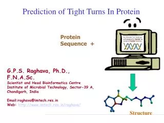
Prediction of Tight Turns In Protein Protein Sequence + G.P.S. Raghava, Ph.D., F.N.A.Sc. Scientist and Head Bioinformatics Centre Institute of Microbial Technology, Sector-39 A, Chandigarh, India Email:raghava@imtech.res.in Web: http://www.imtech.res.in/raghava/ Structure


Prediction of Tight Turns In Protein
E N D
Presentation Transcript
Prediction of Tight Turns In Protein Protein Sequence + G.P.S. Raghava, Ph.D., F.N.A.Sc. Scientist and Head Bioinformatics Centre Institute of Microbial Technology, Sector-39 A, Chandigarh, India Email:raghava@imtech.res.in Web: http://www.imtech.res.in/raghava/ Structure
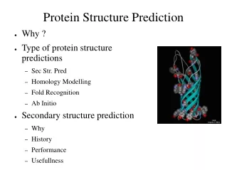
Protein Structure Prediction • Experimental Techniques • X-ray Crystallography • NMR • Limitations of Current Experimental Techniques • Protein DataBank (PDB) -> 27000 protein structures • SwissProt -> 100,000 proteins • Non-Redudant (NR) -> 1,000,000 proteins • Importance of Structure Prediction • Fill gap between known sequence and structures • Protein Engg. To alter function of a protein • Rational Drug Design
Techniques of Structure Prediction • Computer simulation based on energy calculation • Based on physio-chemical principles • Thermodynamic equilibrium with a minimum free energy • Global minimum free energy of protein surface • Knowledge Based approaches • Homology Based Approach • Threading Protein Sequence • Hierarchical Methods • Prediction of intermediate state (Secondary Structure) • Secondary to tertiary structure
Energy Minimization Techniques Energy Minimization based methods in their pure form, make no priori assumptions and attempt to locate global minma. • Static Minimization Methods • Classical many potential-potential can be construted • Assume that atoms in protein is in static form • Problems(large number of variables & minima and validity of potentials) • Dynamical Minimization Methods • Motions of atoms also considered • Monte Carlo simulation (stochastics in nature, time is not cosider) • Molecular Dynamics (time, quantum mechanical, classical equ.) • Limitations • large number of degree of freedom,CPU power not adequate • Interaction potential is not good enough to model
Knowledge Based Approaches • Homology Modelling • Need homologues of known protein structure • Backbone modelling • Side chain modelling • Fail in absence of homology • Threading Based Methods • New way of fold recognition • Sequence is tried to fit in known structures • Motif recognition • Loop & Side chain modelling • Fail in absence of known example
Hierarcial Methods Intermidiate structures are predicted, instead of predicting tertiary structure of protein from amino acids sequence • Prediction of backbone structure • Secondary structure (helix, sheet,coil) • Beta Turn Prediction • Super-secondary structure • Tertiary structure prediction • Limitation Accuracy is only 75-80 % Only three state prediction
Levels of Description of Structural Complexity • Primary Structure (AA sequence) • Secondary Structure • Spatial arrangement of a polypeptide’s backbone atoms without regard to side-chain conformations • , , coil, turns (Venkatachalam, 1968) • Super-Secondary Structure • , , /, + (Rao and Rassman, 1973) • Tertiary Structure • 3-D structure of an entire polypeptide • Quarternary Structure • Spatial arrangement of subunits (2 or more polypeptide chains)
Protein Secondary Structure Secondary Structure Regular Secondary Structure (-helices, -sheets) Irregular Secondary Structure (Tight turns, Random coils, bulges)
Definition of -turn A -turn is defined by four consecutive residues i, i+1, i+2 and i+3 that do not form a helix and have a C(i)-C(i+3) distance less than 7Å and the turn lead to reversal in the protein chain. (Richardson, 1981). The conformation of -turn is defined in terms of and of two central residues, i+1 and i+2 and can be classified into different types on the basis of and . i+1 i+2 i H-bond i+3 D <7Å
a b a:Ramachandran plot showing the characteristic region where -sheet and -helices are found. b: Ramachandran plot showing Type I and II turns represented by a vector
Gamma turns • The -turn is the second most characterized and commonly found turn, • after the -turn. • A -turn is defined as 3-residue turn with a hydrogen bond between the • Carbonyl oxygen of residue i and the hydrogen of the amide group of • residue i+2. There are 2 types of -turns: classic and inverse.
Other rare tight turns • -turn:The smallest is a -turn. It involves only two amino acid residues. The intra-turn hydrogen bond for a -turn is formed between the backbone NH(i) and the backbone CO(i+1). • -turn:An -turn involves five amino acid residues where the distance between C(i) and C(i+4) is less than 7Å and the pentapeptide chain is not a helical conformation. • -turn:The largest tight turn is a -turn, which involves six amino acid residues.
Prediction of tight turns • Prediction of -turns • Prediction of -turn types • Prediction of -turns • Prediction of -turns • Use the tight turns information, mainly -turns in tertiary structure prediction of bioactive peptides
Existing -turn prediction methods • Residue Hydrophobicities (Rose, 1978) • Positional Preference Approach • Chou and Fasman Algorithm (Chou and Fasman, 1974; 1979) • Thornton’s Algorithm (Wilmot and Thornton, 1988) • GORBTURN (Wilmot and Thornton, 1990) • 1-4 & 2-3 Correlation Model (Zhang and Chou, 1997) • Sequence Coupled Model (Chou, 1997) • Artificial Neural Network • BTPRED (Shepherd et al., 1999) (http://www.biochem.ucl.ac.uk/bsm/btpred/ )
BetaTPred: Prediction of -turns using statistical methods (http://imtech.res.in/raghava/betatpred/) Harpreet Kaur and G P S Raghava (2002) BetaTPred: Prediction of -turns in a protein using statistical algorithms.Bioinformatics18(3), 498-499.
Text Output Graphical (Frames) output Consensus -turn
We have evaluated the performance of six methods of -turn prediction. All the methods have been tested on a set of 426 non-homologous protein chains. In this study, both threshold dependent (Qtotal, Qpred., Qobs. And MCC) and independent (ROC) measures have been used for evaluation. Harpreet Kaur and G.P.S Raghava (2002) An evaluation of -turn prediction methods.Bioinformatics18(11), 1508-1514. Performance of existing -turn methods
BTEVAL: A web server for evaluation of -turn prediction methods (http://imtech.res.in/raghava/bteval/) Harpreet Kaur and G P S Raghava (2003)BTEVAL: A server for evaluation of -turn prediction methods.Journal of Bioinformatics and Computational Biology(in press).
BTEVAL: A web server for evaluation of -turn prediction methods
BetaTPred2: Prediction of -turns in proteins from multiple alignment using neural network Harpreet Kaur and G P S Raghava (2003) Prediction of -turns in proteins from multiple alignment using neural network. Protein Science 12, 627-634. • Two feed-forward back-propagation networks with a single hidden layer are used where the first sequence-structure network is trained with the multiple sequence alignment in the form of PSI-BLAST generated position specific scoring matrices. • The initial predictions from the first network and PSIPRED predicted secondary structure are used as input to the second sequence-structure network to refine the predictions obtained from the first net. • The final network yields an overall prediction accuracy of 75.5% when tested by seven-fold cross-validation on a set of 426 non-homologous protein chains. The corresponding Qpred., Qobs. and MCC values are 49.8%, 72.3% and 0.43 respectively and are the best among all the previously published -turn prediction methods. A web server BetaTPred2 (http://www.imtech.res.in/raghava/betatpred2/) has been developed based on this approach.
BetaTPred2 prediction results using single sequence and multiple alignment. Harpreet Kaur and G P S Raghava (2003) Prediction of -turns in proteins from multiple alignment using neural network.Protein Science12, 627-634.
BetaTPred2: A web server for prediction of -turns in proteins (http://www.imtech.res.in/raghava/betatpred2/)
Gammapred: A server for prediction of -turns in proteins (http://www.imtech.res.in/raghava/gammapred/) Harpreet Kaur and G P S Raghava (2003) A neural network based method for prediction of -turns in proteins from multiple sequence alignment. Protein Science12, 923-929.
Network architecture for gamma turns Harpreet Kaur and G P S Raghava (2003) A neural network based method for prediction of -turns in proteins from multiple sequence alignment.Protein Science12, 923-929.
BetaTurns: A web server for prediction of -turn types (http://www.imtech.res.in/raghava/betaturns/) Harpreet Kaur and G P S Raghava (2003) Prediction of -turn types in proteins from evolutionary information using neural network. Bioinformatics (In Press)
AlphaPred: A web server for prediction of -turns in proteins (http://www.imtech.res.in/raghava/alphapred/) Harpreet Kaur and G P S Raghava (2003) Prediction of -turns in proteins using PSI-BLAST profiles and secondary structure information.Proteins(in press).
Contribution of -turns in tertiary structure prediction of bioactive peptides • 3D structures of 77 biologically active peptides have been selected from PDB and other databases such as PSST (http://pranag.physics.iisc.ernet.in/psst) and PRF (http://www.genome.ad.jp/) have been selected. • The data set has been restricted to those biologically active peptides that consist of only natural amino acids and are linear with length varying between 9-20 residues.
3 models have been studied for each peptide. The first model has been ( = = 180o). The second model is build up by constructed by taking all the peptide residues in the extended conformation assigning the peptide residues the , angles of the secondary structure states predicted by PSIPRED. The third model has been constructed with , angles corresponding to the secondary states predicted by PSIPRED and -turns predicted by BetaTPred2. Peptide Extended ( = = 180o). PSIPRED + BetaTPred2 PSIPRED Root Mean Square Deviation has been calculated…….
Averaged backbone root mean deviation before and after energy minimization and dynamics simulations.