Download
1 / 42
420 likes | 704 Vues
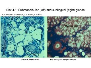
Slot 4.1: Submandibular (left) and sublingual (right) glands. m = mucous, s = serous, x = mixed, d = duct. Mucous acini. Serous demilunes. D = duct; F = adipose cells. Slot 4.2: Parotid Gland. uct. Serous Acini. Slot 4.3: Gastric mucosa diagram (left to right, pyloric, fundic, cardaic).

E N D
Slot 4.1: Submandibular (left) and sublingual (right) glands m = mucous, s = serous, x = mixed, d = duct Mucous acini Serous demilunes D = duct; F = adipose cells
Slot 4.2: Parotid Gland uct Serous Acini
Slot 4.3: Gastric mucosa diagram(left to right, pyloric, fundic, cardaic)
P=parietal C=chief N=mucous neck L=lamina propria Arrows=entero- endocrine cells Slot 4.4: Stomach sections Base of gastric glands Neck of gastric glands P=parietal I=isthmus N=neck L=lamina propria Arrows (left) = mucus Arrows (right) = gastric pits Gastric pits and gastric glands Gastric pits and gastric glands
Slot 4.5: Small intestine sections g = goblet cell; arrowhead = brush border; Arrows = enteroendocrine cells; L = lamina propria; c = capillary, m = smooth muscle cell b = brush border; arrowheads = golgi; Arrows = junctional complexes joining adjacent cells Ileum - villus Intestinal epithelium on villi
Slot 4.7: Large intestine sections Large intestine Colon: Rectoanal junction
Slot 4.6: Appendix L = lumen M = mucosa S = submucosa a = adipose F = muscularis Arrows = crypts Arrowheads = lymph nodules
Slot 4.8: Liver Sinusoids Hepatocytes C = central vein; P = portal canal
LEFT: C = central vein P = portal canal Arrows=sinusoids RIGHT: e = endothelium Arrows=Kupffer cells Arrowhead = binucleate hepa- tocyte Slot 4.10: Liver sections Hepatic artery Bile duct Portal Vein Reticular fibers Sinusoids emptying into central vein = lymph vessel Central Vein Portal Canal
Slot 4.11: Gall bladder sections Simple columnar epithelium Lamina propria Lamina propria c = capillaries a = arteriole v = venule Arrowhead = golgi g = mucous glands M = muscularis A = adventitia
Slot 4.12: Tongue - circumvallate papilla Taste buds Primary papilla Secondary papilla von Ebner’s glands
Slot 4.13: Tongue - foliate papillae Taste buds Secondary papilla von Ebner’s glands
Slot 4.14: Tongue - foliate papillae Secondary papilla Taste buds von Ebner’s glands
Slot 4.16: Esophagus section Mucosa (stratified squamous epithelium) Submucosa Muscularis externa Lamina propria
Slot 4.17: Esophagogastric junction Submucosal mucous glands Muscularis mucosae Gastric pit Note change in epithelial lining at the arrows Stomach = left; Esophagus = right
Slot 4.18: Stomach (fundus and pylorus) Gastric pit Gastric pit Gastric glands Gastric glands Fundus = short pits, long glands Pylorus = deep pits, medium glands
Slot 4.19: Gastric glands Enteroendocrine Cell Chief Cell
Slot 4.20: Gastric glands Parietal Cells Chief Cells
Slot 4.21: Gastroduodenal junction Villi Crypts Brunner’s Glands Note change in thickness of the muscularis externa (Pylorus = right; Duodenum = left)
Slot 4.22: Small intestine sectionsLeft to Right: duodenum, jejunum, ileum Duodenum with Brunner’s glands in submucosa Jejunum with thickest mucosa Ileum with Peyer’s patches in lamina propria/submucosa Peyer’s patches Brunner’s glands
Slot 4.23: Intestinal epithelium (simple columnar) Goblet cell Brush border
Slot 4.24: Duodenum (Brunner’s Glands) Crypt of Lieberkuhn Brunner’s glands
Slot 4.26: Meissner’s plexus/Auerbach’s plexus Muscularis mucosae Muscularis externa Ganglion cells of Meissner’s plexus Ganglion cell of Auerbach’s plexus
Slot 4.28: Ileum Goblet cell Lamina propria Villus
Slot 4.29: Appendix Crypt of Lieberkuhn Lymph nodule Submucosa IC Muscularis externa OL
Slot 4.30: Jejunum (blood supply) Arrow = Vessels run parallel with muscle fibers in inner circular layer of M. externa V = villi S = submucosa F = muscularis externa P = serosa
Slot 4.31: Small Intestine (blood supply) V = villi S = submucosa F = muscularis externa P = serosa V S F P
Slot 4.32: Large intestine section Intestinal crypts – note abundance of goblet cells
Slot 4.33: Liver section Rough outline of liver lobule Portal system Central vein
Slot 4.34: Liver (blood supply) Portal vein Sinusoids Central vein
Slot 4.35: Liver (hepatocytes and sinusoids) Hepatocyte (note binucleate condition) Endothelial cell
Slot 4.36: Liver (portal canals) Hepatic artery Bile duct Lymph vessel Nerve Lymph vessel Portal Vein Bile duct Hepatic artery
Slot 4.37: Liver section Hepatocytes Endothelial cells Central Vein
Slot 4.38: Gall bladder section Simple columnar epithelium Lamina propria Muscularis Adventitia
Slot 4.39: Jejunum Crypts of Lieberkuhn Muscularis mucosae Submucosa Villus Muscularis externa
Slot 2.55: Circumvallate papilla Taste buds Secondary papilla Primary papilla von Ebner’s glands
Slot 2.56: Taste buds (Tongue) Taste buds Taste pore Taste receptor cell Sustentacular cell Basal cell Secondary papilla
Slot 2.59: Fundic stomach Enteroendocrine Cell Chief Cell
Slot 2.60: Duodenum Villus Crypt Brunner’s Glands
Slot 2.61: Ileum Goblet cell Lamina propria Villus