Download
1 / 39
400 likes | 502 Vues
Learn to analyze chest radiographs systematically by examining airway, bones, cardiac area, diaphragm, and lung parenchyma for signs of consolidation, collapse, and interstitial patterns. Understand pulmonary nodules, cardiac anatomy, pulmonary vasculature, kerley A lines, and mediastinal masses for accurate diagnosis.

E N D
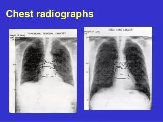
Basics • Anterior-Posterior vs. Posterior-Anterior • AP exaggerates cardiac size • PA requires pt to stand • Look at the whole radiograph • Learn a system - do it the same EVERY time
System • A-B-C-D-E-F • A - Airway/lung fields • B - Bones/soft tissue • C - Cardiac/mediastinum • D - Diaphragm • E - Examine Technique • F - Foreign bodies
Lung Parenchyma • Classify disease into 3 categories • Airspace: alveolar filling • fluffy, opacities, air-bronchograms • Interstitial: lines and small dots • reticulonodular, reticular, nodular • Nodule: single or multiple, vary in size, w/ or w/o cavitation/calcification, smooth or irregular
Consolidation • Filling or loss of air spaces • Pus - Pneumonia • Fluid - Pulmonary edema • Blood - infarct, hemorrhage • Foreign body - aspiration • Tumor - bronchoalveolar carcinoma • Volume loss - atelectasis
Consolidation • Radiographic signs • Opacity, air bronchograms, silhouetting • Silhouette sign: intrathoracic lesion touching border of heart, aorta, diaphragm obliterating that border • Helps to identify location of consolidation
Left Heart Silhouette sign
Consolidation • Silhouette sign: • What structure is silhouetted on PA? • R heart = RML • L heart = lingula • Aorta, diaphragm = Lower lobe • Lateral view: which diaphragm is silhouetted? • Fissure sign: abrupt edge @ margin • Increased density of vert. just above diaphragm on lateral
Collapse • Atelectasis - volume loss • Extrinsic compression (effusion, tumor, etc) • Airway obstruction • Extraluminal - tumor, LAD • Intraluminal - tumor, foreign body • Lobar collapse: mediatstinal shift to affected side, displacement of hilum/fissures, fewer vessels on affected side
Interstitial Pattern • Acute process: • Pneumonia - viral, fungal, Tb, PCP • Edema - CHF, Renal failure w/ overload • Drug/Transfusion reaction • Chronic: many etiologies • Normal/low lung volumes
Interstitial Pattern • Upper lobe predominant • Tb, pneumoconioses, fibrosis from ankylosing spondylitis • Mid lung predominant • sarcoid, berylliosis, allergic alveolitis, eosinophilic granulomatosis • Lower lung predominant • IPF, lymphangitic tumor spread, CVD fibrosis, chronic edema, drug rxn
Interstitial Pattern • Large Lung volumes: indicates air trapping • Cystic fibrosis • Eosinophilic granulomatosis • Lymhangioleiomyomatosis • Tuberous sclerosis
Pulmonary Nodule(s) • Solitary Nodule: many etiologies • Primary lung tumor, mets, granuloma, septic emboli, pulmonary AVM, hamartoma, Wegener’s vasculitis, bronchiectasis, fungal infection, etc • Important features • Change over time: growing is worrisome • Calcification: eccentric is worrisome • Size: > 3cm more worrisome
Pulmonary Nodule(s) • Multiple Nodules • Metastatic until proven otherwise • septic/bland emboli • vasculitides, CVD • pneumoconioses • Eosinophilic granulomatosis • Fungi, viral, Tb PNA • Wegener’s • Lymphoma
Cardiac Anatomy • Frontal view • Right atrium • SVC • Aortic knob • Left atrial appendage • Left ventricle Lateral view Right atrium/Ventricle Left ventricle Left atrium Aortic arch Main Pulm. Artery Descending Thoracic Aorta
Cardiac Anatomy • On frontal CXR - 45% or less than largest diameter from inner aspect of rib to rib laterally • Right heart border - mostly RA • Left Border - Aortic arch, MPA, LAA, LV
Atrial/Ventricular Hypertropy • Right Atrium - Right border >4cm from center of spine • Right Ventricle - fills retrosternal space >1/3 distance between diaphragm & sternomanubrial joint • Left Atrium - subcarinal angle >90 degrees, posterior deviation of left main stem bronchus • Left Ventricle - LV reaches spine prior to diaphragm
Pulmonary Vasculature • Many potential patterns to help narrow differential for cardiac disease • 3 you need to know • Normal - lower lobe vessels larger due to gravity, taper smoothly to periphery, interlobar arterial size (11-16mm M, 9-14mm F)
Pulmonary Vasculature • Pulmonary venous hypertension: upper lobe vessels larger “cephalization” result of hypoxic vasoconstriction; dependent edema • LV failure (ASCHD, valvular), atrial myxoma, PVOD • Pulmonary arterial hypertension: “pruning” or rapid tapering of peripheral vessels from large central arteries • Chronic venous HTN, COPD, Chronic PE, vasculitides, Primary PHTN, L-to-R shunt
Mediastinum • Several compartments • Anterior: ant. = sternum, post. = pericardium • Middle: ant. = pericardium, post. = trachea • Posterior: ant. = trachea, post. = ribs • Don’t miss a widened mediastinum = could be an aortic aneurysm
Mediastinum • Masses by compartment • Anterior: “4T’s” • Teratoma • Thymoma • Terrible tumor (lymphoma, mets) • Thyroid - goiter • Middle: • Aortic aneurysm
Mediastinum • Lymph nodes - Lymphoma/Mets • Pericardial/bronchogenic cyst • Posterior: • Aneurysm • Lymph nodes • Neurogenic tumors - ganglion tumor • Spine - osteophyte • Esophagus - paraesophageal hernia • Substernal Thyroid
Pleural Abnormalities • Effusions: fluid • 300-500cc to blunt CP angle on frontal • 150cc posterior to blunt CP angle on lateral • Free flowing or not?: obtain bilateral decubital films • Subpulmonic: lateral peaking of diaphragm, loss lung parenchyma below diaphragm
Pleural Abnormalities • Pneumothorax: air in pleural space • Apical or “deep sulcus” • Tension: flattened ipsilateral lung on mediastinum • Masses • Angle w/ chest wall is obtuse • Center of Mass • Well defined margin only on 1 side
Pleural Abnormalities • Thickening • Focal: unilateral • usually from infection/hemorrhage • Plaque from asbestosis - near • diaphragms • Diffuse: unilateral • Smooth: Old Tb, empyem, hemothorax, • mesothelioma, mets, lymphoma • Nodular: same except Tb