Download
1 / 58
580 likes | 910 Vues
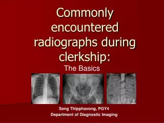
Commonly encountered radiographs during clerkship:. The Basics. Seng Thipphavong, PGY4 Department of Diagnostic Imaging. Objectives and Outline. To review the commonly encountered radiographs during clerkship, with a review of radiographic anatomy and disease entities Radiographs:

E N D
Commonly encountered radiographs during clerkship: The Basics Seng Thipphavong, PGY4 Department of Diagnostic Imaging
Objectives and Outline • To review the commonly encountered radiographs during clerkship, with a review of radiographic anatomy and disease entities • Radiographs: • The Chest Radiograph • The Abdominal Radiograph • Miscellaneous Radiographs…
The Chest Radiograph Anatomy Cases (3)
Anatomy trachea clavicle aortic arch SVC aortopulmonary window main pulmonary artery left atrial appendage right atrium left ventricle right hemidiaphragm left hemidiaphragm
Anatomy trachea retrosternal airspace left pulmonary artery right pulmonary artery right heart chambers left heart chambers IVC
Case 1 • 69 y.o. female presents with shortness of breath
Case 1 Kerley B lines peribronchial cuffing
Pulmonary edema • Radiographic signs of pulmonary edema? (5) • Enlarged cardiac silhouette • Kerley B lines (fluid in the interlobular septae) • Peribronchial cuffing • Indistinctness of the pulmonary vessels • Pleural effusion
Case 2 • 69 y.o. with fever and cough
Case 2 Air bronchograms
Case 2 • Findings of pneumonia on radiograph? • Consolidation (white) and air bronchograms • How are pneumonia and atelectasis similar on radiograph? • Both are white • How are pneumonia and atelectasis different on radiograph? • Look for air bronchograms • Atelectasis will have signs of volume loss
Case 3 • 69 y.o. with chest pain
Case 3 Visceral pleura
Case 3 • Causes of pneumothorax? • Numerous! • Treatment? • Urgent • Chest tube • 25 G needle 2nd intercostal space
Case 3 • Deep sulcus sign? • pneumothorax on supine films • especially seen in ICU patients
The Abdominal Radiograph Anatomy Cases (3)
Anatomy Right kidney Left kidney Hepatic angle Left psoas Properitoneal fat Air in descending colon
Case 1 • 69 y.o. with abdominal pain
Case 1 • What films are obtained in a conventional abdominal series? • Supine and upright abdomen, chest radiograph • What are the 4 cardinal symptoms of small bowel obstruction? • Nausea, vomiting, abdominal distension, obstipation • What are the causes of SBO? • Adhesions, hernia, stricture, neoplasm, gallstone ileus
Case 1 • What are the signs of SBO on radiograph? • Dilated and fluid filled loops, “step-ladder” appearance • What is the difference between ileus and SBO? • SBO indicates mechanical obstruction • Ileus is an adynamic state (“bowel shuts down”)
Case 2 • 69 y.o. with abdominal pain
Case 2 Cupola sign Football sign
Case 2 • Signs of free intraperitoneal air on upright radiograph? • Air under the diaphragm • Signs of free intraperitoneal air on supine radiograph? • “football sign”, football shaped lucency central abdomen • “cupola sign”, free air in the mid-subphrenic space • What is Rigler’s sign? • Free air outlining both sides of bowel
Companion case Rigler’s sign
Case 2 • What are the 2 most common reasons to see free intraperitoneal air? • Post-operative or perforated duodenal ulcer • Is free air commonly seen on radiograph from perforated diverticulitis? • No. • Why? • the omenteum usually contains the air, and is not seen on radiograph
Case 3 • 69 y.o. with abdominal pain
Case 3 • What are the signs of large bowel obstruction? • Dilated large bowel proximal to the site of obstruction • Paucity of air distal to obstruction • What are the most common causes of large bowel obstruction? • Colon Ca, stricture (post-inflammatory diverticulitis or IBD), volvulus
The Miscellaneous Radiograph Cases (4)
Case 1 • 69 y.o. in a fight
Case 1 • What is a Boxer’s fracture? • Fracture of the 5th metacarpal • Potential complications of a Boxer’s fracture? • Metacarpal shortening • Usually the distal fragment is rotated in a radial direction, and may heal with deformity
Wrist and hand anatomy Distal phalynx DIP joint Middle phalynx PIP joint Proximal phalynx MCP joint Sesamoid Metacarpal CMC joint Distal ulna Distal radius
Wrist anatomy hamate trapezoid capitate trapezium pisiform triquetrum scaphoid lunate
Case 2 • 69 y.o. who fell
Case 2 • What is the classic clinical presentation for a hip fracture? • Shortened lower extremity and external rotation
Pelvic anatomy Iliac crest SI joint Sacral ala Femoral head Iliopectineal line Greater trochanter Femoral neck Superior pubic ramus Ischial tuberosity Lesser trochanter Obturator foramen Inferior pubic ramus Pubic symphysis
Case 3 • 69 y.o. who fell