Download

1 / 36
540 likes | 1.37k Vues
Regulation of blood flow. DR. Shafali Singh. Learning objectives. Define autoregulation of blood flow. Describe how the theory of metabolic regulation of blood flow accounts for active hyperemia and reactive hyperemia.

E N D
Regulation of blood flow DR. Shafali Singh
Learning objectives • Define autoregulation of blood flow. Describe how the theory of metabolic regulation of blood flow accounts for active hyperemia and reactive hyperemia. • Describe the contribution of myogenic tone to blood flow regulation. • Identify the role of PO2 , PCO2 , pH, adenosine, and K+ in the metabolic control of blood flow to specific tissues. • Diagram the synthetic pathway for nitric oxide (EDRF, endothelial derived relaxing factor), including substrate and the interplay between endothelium and vascular smooth muscle.
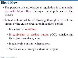
Microcirculation & Vasomotion • Circulation of blood through the smallest vessels of the body-arterioles, capillaries, and venules. • The rhythmic oscillatory behavior of capillaries is caused by contraction and relaxation (vasomotion) of the precapillary vessels (i.e., the arterioles and small arteries
An increase in transmural pressure, caused either by an increase in venous pressure or by dilation of arterioles, results in contraction of the terminal arterioles. A decrease in transmural pressure causes precapillary vessel relaxation . • Humoral and possibly neural factors also affect vasomotion
Transcapillary Exchange • Solvent and solute move across the capillary endothelial wall by three processes: • Diffusion, • Filtration, • Pinocytosis
For diffusion across a capillary wall, Fick's law can also be expressed as
Local Control of Blood Flow: • There are several examples of local control of blood flow, including: • Autoregulation • Active hyperemia • Reactive hyperemia
Active hyperemia : blood flow to an organ is proportional to its metabolic activity. • When any tissue becomes highly active e.g • An exercising muscle, • A gastrointestinal gland during a hypersecretory period, • Brain during rapid mental activity, • the rate of blood flow through the tissue increases.
Reactive hyperemia : increase in blood flow in response to or reacting to a prior period of decreased blood flow (arterial occlusion).
MECHANISMS FOR CONTROL OF REGIONAL BLOOD FLOW INTRINSIC EXTRINSIC Autoregulation Autonomic Nervous system Myogenic Metabolic
Intrinsic Regulation (Autoregulation) • The control mechanisms regulating the arteriolar smooth muscle are entirely within the organ itself. • No nerves or circulating substances are involved in autoregulation. • Thus, the autonomic nervous system and circulating epinephrine have nothing to do with autoregulation. • There are 2 main theories that attempt to explain autoregulation. Of them, the metabolic hypothesis has more support.
Two basic mechanisms are proposed to explain the phenomena of autoregulation and active and reactive hyperemia: Metabolic hypothesis. Myogenic hypothesis
Intrinsic Regulation (Autoregulation) Metabolic hypothesis • Tissue produces a vasodilatory metabolite that regulates flow, e.g., adenosine in the coronary circulation • A dilation of the arterioles is produced when the concentration of these metabolites increases in the tissue. • The arterioles constrict if the tissue concentration decreases.
Vasodilator Theory for Acute Local Blood Flow Regulation— • Adenosine • Carbon dioxide, • Adenosine phosphate compounds, • Histamine • Potassium ions, • Hydrogen ions.
Autoregulation is the maintenance of a constant blood flow to an organ in the face of changing arterial pressure. Several organs exhibit autoregulation of blood flow, including the kidneys, brain, heart, and skeletal muscle. If BP fallscompensatory vasodilation of the coronary arterioles decreases the resistance of the coronary vasculature flow is maintained constant .
Intrinsic Regulation (Autoregulation) Myogenichypothesis • Increased perfusing pressure causes stretch of the arteriolar wall and the surrounding smooth muscle. • Because an inherent property of the smooth muscle is to contract when stretched, the arteriole radius decreases, and flow does not increase significantly.
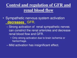
Blood flow in most cases is proportional to tissue metabolism. • Blood flow is independent of nervous reflexes (e.g., carotid sinus) or circulating humoral factors. • Autoregulating tissues include (tissues least affected by nervous reflexes): Cerebral circulation Coronary circulation Skeletal muscle vasculature during exercise • Control of renal blood flow is also commonly referred to as autoregulationeven though it is partially controlled by neural and hormonal influences
Extrinsic Regulation • These tissues are controlled by nervous and humoral factors originating outside the organ, e.g., resting skeletal muscle.
The main mechanism controlling flow in resting skeletal muscle and all other major extrinsically regulated systemic circuits is tonic changes in sympathetic adrenergic activity, i.e., norepinephrine acting on α receptors, causing constriction. Generally, the parasympathetic system does not affect arterioles, and thus it has little or no influence on TPR (exception: the penis). Circulations with mainly extrinsic regulation (those most affected by nervous reflexes): Cutaneouscirculation Resting skeletal muscle
Control of Resting versus Exercising Muscle Resting muscle • Flow is controlled mainly by increasing or decreasing sympathetic α -adrenergic activity. But β2 receptors can contribute to the regulation of blood flow. Exercising muscle • The increase in flow is mainly via vasodilatory metabolites, but this cannot occur without a significant contribution via an increase in CO. • β2 activation via circulating epinephrine can contribute to the increase in flow. • Sympathetic adrenergic nerves have no effect on flow in exercising muscle. (α receptors become less responsive to norepinephrine.) • Thus, if there was an increase in sympathetic activity to an exercising muscle, flow would not be altered significantly by α receptor-mediated effects.
Muscle Blood Flow. ml/100 g Muscle/min • Resting blood flow 3.6 • Blood flow during maximal exercise 90
Increase in flow results from intra-muscular vasodilation caused by the direct effects of increased muscle metabolism. • Moderate increase in arterial blood pressure that occurs in exercise, usually about a 30 per cent increase. • The increase in pressure not only forces more blood through the blood vessels but also stretches the walls of the arterioles and further reduces the vascular resistance.
Exercising skeletal muscle • Blood flow increases. • Vascular resistance decreases. • Capillary pressure increases. • Capillary filtration increases. • Lymph flow increases. • Venous PO2decreases and can reach extremely low levels. • Extraction of oxygen increases.
Pulmonary response to exercise • A large increase in cardiac output means increased volume pumped into the circuit. • This will produce a rise in pulmonary pressures. • Because of the passive, compliant nature of the circuit, the response to a rise in pressure is vessel dilation. • This response leads to apical blood vessel recruitment. • The overall response is a large decrease in resistance.
Pulmonary Circuit • Blood flow (CO): large increase • Pulmonary arterial pressure: slight increase • Pulmonary vascular resistance: large decrease • Pulmonary blood volume: increase • Number of perfused capillaries: increase • Capillary surface area: increase, i.e., increased rate of gas exchange
Q Correct statements about the increase in pulmonary blood flow during vigorous exercise include which of the following? a. The percentage of increase in flow is greater in the bases of the lungs than in the apices b. The increase in flow is caused by a greater-than-fivefold increase in pulmonary arterial pressure c. The increase in pulmonary blood flow is less than the increase in systemic blood flow d. The increase in pulmonary blood flow is accommodated by dilation of pulmonary arterioles and capillaries e. The increase in pulmonary blood flow is caused by sympathetic nerve stimulation of the pulmonary vasculature
Regional Circulations • Blood flow to active organs like Heart, Skeletal muscle. • Blood flow to inactive organs like Kidney, Skin, GIT
Regional Circulations • Cutaneous blood flow Initial decrease, then an increase to dissipate heat • Coronary blood flow Increase due to increase in volume work of the heart • Cerebral blood flow No significant change (arterial CO2 remains unchanged) • Renal and GI blood flow Any change would be a decrease. This is more likely in the splanchnic circuit.