Download
1 / 21
240 likes | 833 Vues
A Tour of the Cell. (Chapter 6). The Discovery of the Cell: Hooke. In 1665, Robert Hooke used an early compound microscope to look at a thin slice of cork, which is a plant material. He noticed that cork looked like thousands of tiny, empty chambers which he called “cells”

E N D
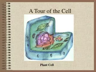
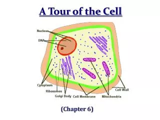
A Tour of the Cell (Chapter 6)
The Discovery of the Cell:Hooke • In 1665, Robert Hooke used an early compound microscope to look at a thin slice of cork, which is a plant material. • He noticed that cork looked like thousands of tiny, empty chambers which he called “cells” • We now know cells are the basic units of life.
The Discovery of the Cell:Von Leeuwenhoek • The existence of cells was unknown for many years, but this changed with the invention of the microscope. • Anton von Leeuwenhoek used a single-lens microscope to observe pond water and other things. • The microscope revealed a world of tiny living organisms.
Developing the Cell Theory • In 1838, Matthias Schleiden concluded that all plants were made of cells. • In 1839, Theodor Schwann stated that all animals were made of cells. • In 1855, Rudolph Virchow concluded that new cells were created only from division of pre-existing cells. • These discoveries led to the cell theory.
Compound Light Microscope • Combination of lenses and light used to magnify small objects held on a slide • Live specimen may be observed (ie, pond water) • Max magnification = 1000x
Electron Microscopes • Image produced on a computer screen using a beam of electrons rather than light • More powerful (300,000x or more) than light microscopes, but specimen cannot be alive • Transmission • Study of inner structure of a specimen • Samples are cut into thin slices for viewing • Images are 2-D • Scanning • Allows study of specimen surface • Images are 3-D
The Cell Theory • All living things are composed of cells. • Cells are the basic units of structure and function in living things. • New cells are produced from pre-existing cells.
Self-Assessment • What are the three key ideas of the cell theory? • What scientists contributed to the development of the cell theory? How did they contribute? • Identify the type of microscope most useful for viewing each of the following: • A group of cells in a thin layer of onion skin • The details of the surface of a human hair • The detailed structure of a mitochondria inside a muscle cell
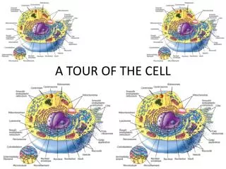
Two Types of Cells • All cells are surrounded by a barrier called a cell membrane and contain DNA • Eukaryotic cells contain a nucleus & membrane organelles. (plants, animals, fungi & protists.) • Prokaryotic cells do NOT contain a nucleus (still have DNA) and most organelles (do have ribsomes) and are classified as bacteria.
Cell Boundaries • All cells are surrounded by a thin, flexible barrier, the plasma or cell membrane, which acts as a gate-keeper, regulating what enters and leaves the cell, and also provides some protection and support • Many cells also produce a strong supporting layer around the membrane known as a cell wall. The main function of the cell wall is support and protection for the cell.
Structure of the Cell Membrane • The composition of nearly all cell membranes is a double-layered sheet called a lipid bilayer. Lipid bilayer
Functions of the Plasma Membrane • Embedded enzyme proteins help carry out chemical reactions of the cell • Receptor proteins allow to receive chemical messages • Surface molecules allow recognition & communication between cells • Transport proteins serve as channels or pumps to move materials in and out of cells • Proteins from adjacent cells allow intracellular joining
Diffusion Through Cell Boundaries • Particles in a solution tend to move from an area where they are more concentrated to an area where they are less concentrated.(visualize this as a downhill movement, [high] to [low]) • This process is called diffusion • When the concentration of the solute is the same throughout a system, the system has reached equilibrium.
Osmosis • The diffusion of water through a selectively permeable membrane.
Osmotic Pressure & Tonicity • If you compare two solutions, the more concentrated solution (less water) ishypertonic. • The more dilute (more water) solution is hypotonic. • When the concentration is equal in both solutions, the solution is described as isotonic.
Facilitated Diffusion • Cell membranes have protein channels that act as channels (pores) or carrier molecules, making it easy for certain molecules to cross. • The movement of specific molecules across cell membranes through protein channels is known as facilitated diffusion. • This process usually involves solute molecules (rather than solvent molecules) and is a form of passive transport because it requires no energy, ([high] to [low])
Active Transport • Active transport involves the use of energy (ATP) to move substances across a cell membrane • Ex) Movement of materials in the opposite direction from which the materials would normally move - against a concentration difference • This is achieved by membrane proteins that act as pumps • Visualize this as UPHILL, [low] to [high]) • Large materials also move by active transport regardless of concentration gradient
Endocytosis is the process of taking large materials into the cell using vesicles, or pockets, of the cell membrane.(endo=enter) Phagocytosis – cell “eating”; actively moving solids into the cell Pinocytosis – cell “drinking”; actively moving liquids into the cell Exocytosis, the membrane of the vesicle surrounding the material fuses with the cell membrane, forcing the contents out of the cell (exo=exit) Active Transport of Large Materials(“Bulk Transport”)
Summary Assessment • What is the difference between active and passive transport? • Name and describe three examples of each. • Distinguish between hypotonic, hypertonic and isotonic solutions. • Explain what will happen to an animal cell in each. • Explain what will happen to a plant cell in each.