Download
1 / 1
20 likes | 180 Vues
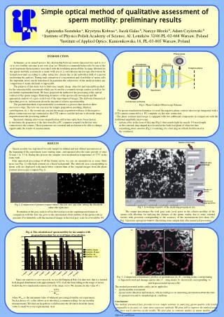
Fig..4. The calculation of sperm motility for ten samples with proposed procedure for several times of incubation.

E N D
Fig..4. The calculation of sperm motility for ten samples with proposed procedure for several times of incubation • Since our statistics is not very rich, we use the Empirical Rule for data sets: that is a normal, bell-shaped distribution with approximately 95% of all the data falling in the range of errors • bellow the two standard deviation (2) of the mean (<x>). We assume for the value of c: • where Wmaxis the maximum value of whiteout percentage found in our experiment. • Such a choice of c value allows as to introduce a common scaling for our motility • measurements. We find our hypothesis valid because the deviation from the mean • value is small for every experimental trial. Simple optical method of qualitative assessment of sperm motility: preliminary results Agnieszka Sozańska a, Krystyna Kolwas a, Jacek Galas b, Narcyz Błocki b, Adam Czyżewski b a Institute of Physics Polish Academy of Science, Al. Lotników 32/46 PL-02-668 Warsaw, Poland bInstitute of Applied Optics, Kamionkowska 18, PL-03-805 Warsaw, Poland • INTRODUCTION • In humans, as in animal species, the relationship between semen characteristics and in vivo or in vitro fertility outcome is not very clear yet. Motility is commonly believed to be one of the most important characteristics associated with the fertilizing sperm ability. In many laboratories the sperm mobility assessment is made with use of a conventional microscope observation by trained personnel according to rather subjective criteria due to the individual skill of a person performing the analysis. During such estimation of concentration and of mobility of sperm cells the important errors can be introduced. In particular the subjectivity of the analysis makes any comparison of results difficult or impossible. • The purpose of this study was to find some simple, cheap, objective and repeatable method • for the semen motility assessment which can be used in a common storage centres as well as for • our further experimental trials. We have proposed the method of the processing of the optical • contrast of the sperm images illustrating dynamics of the sperm cells movement and the • appropriate analysis of a grey scale level of the superimposed images. The elaborated numerical • algorithm gives us information about the amount of relative sperm motility. • The presented method of sperm motility assessment is a process that involves three • successive steps. The first one concerns the sample preparation (washing, dilution, • centrifugation, etc.); the second one concern the image acquisition with use of the negative • phase-contrast microscope connected to the CCD camera; and the last one is about the image • acquisition and the processing method. • Specimen staining, microscope magnification and system optics have been chosen • to maximise the properties of the data stored by a PC computer coupled via the fire-wire • connection to the camera. Those parameters are essential and are known to be able to change • significantly the results of measurements. Fig.1. Phase Contrast Microscope Scheme • For sperm visualization dynamics we used the negative phase contrast microscope integrated with a CCD camera connected to a PC computer via fire wire connection. • The phase contrast microscope is equipped with two additional components in comparison with the traditional amplitude microscope: • a phase plate in the form of the ring (Fig.1) that retards light by exactly 1/4 wavelength • in the centered, ring-shaped area located at the back foal plane of objective lens • a matching phase annulus (Fig.1) consisting of a clear ring on a black field located in • the condenser RESULTS Sperm motility was registered for each sample for diluted and non diluted spermatozoa at the beginning of the experiment (zero starting time), and repeated after the same periods of time: 30 min, 1 h, 1.5 h. During this process the samples were incubated in temperature of 37°C in the water bath. After numerical processing of all the frames movie we can see spermatozoa as some white spots (see Fig. 2) with high contrast on a black background. The whiteout area corresponding to sperm cells are displayed with much better contrast than at the original images from the phase contrast microscope (compare Fig.2). Fig.2. Comparison of pictures frames before (the left one) and after setting the threshold value (the right one). Fig.3. A working window of the analysing program in use. The output percentages the total gray scale level gives us the relative motility of the sperm cells allowing for studying the changes of the sperm vitality due to some external factors with precision corresponding to the accuracy of the measurement (less than 1%). Fig. 3 presents a program window illustrating some sample data after numerical processing. Evaluation of the gray scale level for the total area of the superimposed frames in comparison with the first one gives as the information about motility of the sperm cells in percents. For immobile cells the measured change in the total gray scale level would be 0%. A B C D Fig. 5. Comparison of kinematics profiles of spermatozoa (A, B – circling tracks corresponding to hyperactivated and damage sperm cells, C – long tracks, D- ideal tracks corresponding to non-hyperactivated sperm cells). • The method presented in this study can be applied to: • sperm motility assessment • sperm tracks detection and analysis, which could gives us interesting information about the state • of spermatozoa and its morphological condition. • Conclusions: • The method presented here provides a new simple solution in analyzing sperm quality with results comparable in accuracy to some more expensive methods. We plan still to improve the method using still more reach statistics of the results. We also plan to continue studies of sperm motility under different conditions.