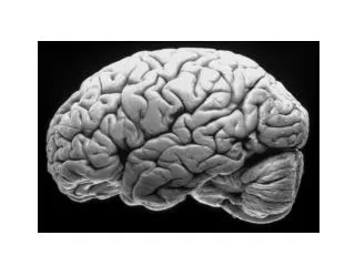
Visual Cortex
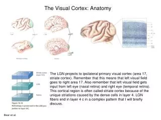
Visual Cortex. • A hemisphere registers the contralateral visual field. About half of the LGN and over half of the visual cortex is devoted to the fovea, so macular sparing occurs with loss of cortex because the fovea’s area is so big that you usually don’t lose it all.

Visual Cortex
E N D
Presentation Transcript
• A hemisphere registers the contralateral visual field. • About half of the LGN and over half of the visual cortex is devoted to the fovea, so macular sparing occurs with loss of cortex because the fovea’s area is so big that you usually don’t lose it all.
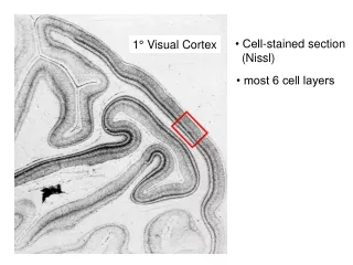
• V1 (aka striate cortex) is found in the banks of the calcarinesulcus, and it has three functions: • 1. Begins to process incoming visual info and put together parts of the visual scene. • 2. It directs information to the appropriate subsequent location.
3. It provides a spatially accurate template. • • There is a retinotopic organization in V1. • The fovea is caudal. • The upper visual field is in the lower bank of the calcarinesulcus. • The lower visual field is in the upper bank.
There is also a temporal crescent, which is the part of the visual field only seen by one eye. • • In V1, inputs from the LGN remain segregated so that they can be combined in an orderly way.
The layers of V1 allow for serial processing and increasing complexity. • Layer 4 in V1 is particularly wide and complex. M layers of the LGN project to layer 4Cα, whereas P layers of the LGN project to layer 4Cβ. • This organization and thickness of layers changes at the border with V2.
• Even at the first synapse in V1, receptive fields begin to become more complex. • Neurons in layer 4 of V1 respond to bars (light or dark) of particular orientations. • The cells that respond to bars like this are called simple cells.
The orientation sensitivity of cells changes in an orderly manner as you move laterally throughout the cortex. • All neurons in a column respond to the same orientation.
• You get bars by taking input from multiple center/surround ganglion cells that line up so their RFs overlap to form a bar. • This is known as the convergence of excitation model. • Some of the same LGN cells can contribute to overlapping bars and project to multiple V1 neurons for multiple orientations.
• In addition to layers, as you move laterally you see that the primary visual cortex is also organized into columns. • The cells directly above and below each other are sensitive to the same orientation of bars. • Moving 50 microns will bring you to another column, different by about 10 degrees. • So 1mm of V1 encompasses around 180 degrees of rotation.
• There are also ocular dominance columns, which are arranged in alternating slabs across the surface of the cortex. • The inputs from each eye reach the cortex separately (layers 2, 3, and 5 go to one column, and layers 1,4, and 6 would go to an adjacent column).
These columns are about 1mm wide. • Though at other layers the signals from the two eyes converge, neurons in a column respond preferentially to the eye that directly innervates layer 4 at that location.
• Ocular dominance columns are present with a fair amount of overlap in layer 4 at birth. • Over about 5 years, tight borders between R and L eye regions are established in layer 4.
This is a sensitive time in visual development. If you deprive one eye of vision, its territory is taken over by the good eye (though this eye sees no better).
This can be treated by a patch that forces the deprived eye to be more active. • This results in a pattern of activity also seen in strabismus (deviation of eyes from center point), in which all 6 layers of V1 are driven only by 1 eye.
The size of left and right eye columns becomes normal, but no neurons in any layer are driven by both eyes. • • Seeing in depth (stereopsis) requires V1 neurons that are responsive to input from both eyes. • This is absent if you fix a depraved-eye with a patch.
Eye specific inputs remain segregated in layer 4, but they combine in layer 3 to produce steropsis. • Layer 4 is the last place you see neurons driven only by 1 eye.
• So far, what we’ve talked about applies to inputs from the M and P cells. • K layers of the LGN (for blue-on cells) project to patches of layer 3 in V1. • These are called blobs.
• A block, or ice cube, contains two ocular dominance columns (one R, one L) each with blobs .5 mm apart lined up at their centers, as well as 180 degrees worth of orientation columns running perpendicular to the ocular dominance columns.
So one ice-cube has the machinery to process all types of visual info. • • M retinal ganglion cells → magnocellular LGN → V1 Layer 4Cα → V1 Layer 4B → V2 Thick stripes → Middle Temporal Cortex (aka V5) → Dorsal stream, responds to 3D aspects of vision, especially movement and telling if you’re moving or something else is moving.
o Layer 4Cα also contributes to the blobs. • • P retinal ganglion cells → parvocellular LGN → V1 Layer 4Cβ → V1 interblobs → V2 interstripes → V4 → Ventral stream, carries info about shape, like people’s faces.
o Layer 4Cβ also contributes to the blobs • • Blue-On ganglion cells → koniocellular LGN → blobs (get input from all 3 ganglion cell types) → Ventral stream, carries info about color.