Download
1 / 1
10 likes | 1.05k Vues
Determining Titer of M13 Bacteriophage in an Unknown Stock Solution Ms. Sue Barnstuble-Kingman, Costa Mesa High School, High School Biology and Life Science Teacher UCI Summer Biotechnology Institute July 19-23, 2004 Objective

E N D
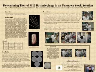
Determining Titer of M13 Bacteriophage in an Unknown Stock Solution Ms. Sue Barnstuble-Kingman, Costa Mesa High School, High School Biology and Life Science Teacher UCI Summer Biotechnology Institute July 19-23, 2004 Objective To determine the titer, or concentration of a virus (filamentous bacteriophage M13mp18), in a stock solution using serial dilutions. • Procedure • Perform serial dilutions of a stock solution of bacteriophage M13mp18 so that dilutions at 10–5, 10-7, 10-9, and 10-11 are made. Dilute the virus concentration using 990µL of Phosphate Buffer Saline (PBS). This is done because there are usually too many viruses in a stock solution, and the whole plate will become plaqued, making it impossible to differentiate between infected and uninfected areas. Because of this, serial dilutions are performed to ensure that there aren’t too many viruses in the solution and not enough bacteria. Each virus needs to have a bacterium that it can infect. This also lets us know what the most effective dilution is for counting and distinguishing plaques from uninfected areas of agar. • Dispense one drop of E. coli bacteria into each of four test tubes, and then transfer 100µL of each bacteriophage dilution into each tube. • After incubating overnight to let the viruses infect the bacteria, and allow the bacteria to replicate, count the number of plaques that appear on each plate to determine the titer, or viral concentration, of the original stock solution. Each plaque is a group of bacteria that have been infected and are, therefore, growing at a slower rate than the bacteria around them. Background Viruses are parasites of cells. They attack, kill and replicate. Viruses that infect bacteria are known as bacteriophages. There are many different types of viruses, they are very specific about the organisms or types of cells they attack, and they have different means of infection. M13 is a filamentous (long and thin) bacteriophage that attacks E. coli bacteria and is important in genetic engineering experiments. It attaches to sites on the outside of bacteria known as pili. After attaching to these sites, the virus is able to enter the cell, remove its protective coating, and release its DNA into the bacterium. Soon, the virus has taken over the bacterium and gets the cell to make viral DNA and the proteins it codes for. M13 does not kill the cell, but the cell stays infected, and because it is producing more viral DNA, the bacterium does not grow as fast as normal. The cells around this infected bacterium also become infected, which can be seen when looking at an infection on an agar plate or Petri dish. The infected bacteria appear as lighter patches since they are growing slower than their neighbors. These lighter areas are known as plaques, or Plaque Forming Units (PFU). By counting PFU, it is possible to determine the concentration of infectious virus, also known as titer, in a solution Using micropipettes to transfer very accurate amounts of stock solution and PBS for serial dilutions M13 Bacteriophage (virus) used Artist’s Representation by Russell Knightley media Adding E. coli bacteria to the dilutions Results Labeling top agar before liquifying, then cooling it to prepare it for bacteria and virus solution Using sterile technique to transfer the solution to the agar plates Putting agar plates in incubator overnight Extension After performing this experiment and a bacterial transformation, it appears that the viral infection is much more effective than the bacterial transformation at making a bacteria produce a new type of DNA and new set of proteins. A virus is 100% effective in transferring DNA into a bacterium, while the plasmids tested were only 0.05% in transferring their DNA into the bacteria. E.coli bacteria cell Pili are the structures on the outside of the cell that the virus attaches to and that the bacterium uses for asexual reproduction. Artist’s Representation by Russell Knightley media Conclusion After looking at the plates with different serial dilutions of an unknown bacteriophage concentration in a stock solution, I determined the dilution of 10-9 was the best for counting plaques and calculating the titer of the original stock. The dilutions of higher concentration contained too many plaques to count, they all ran together to form one across the agar plate. The plaques, or spots, show bacteria that are growing slower than their neighbors because they are infected with M13 bacteriophage. By counting the plaque forming units (PFU), one can determine the titer, or concentration of virus, in the stock solution. Since there were 162 PFU/0.1mL in a 10-9 dilution, the concentration of the stock would be: 162 PFU/ 0.1 mL x 1/10-9 = 162 x 1010 = 1.62 x 10¹² PFU/mL This number is verified by the calculated titer based on the number of PFU observed in a dilution of 10-11, so it is a valid calculation. Based on these calculations, I determined that dilution of 10-7 would have 16,500 PFU (too many to count), and a dilution of 10-5 would have 1,6500,000 PFU (even more to count!). Adding the bacteria/plasmid solution to the agar plate for bacterial transformation follow-up experiment Spreading bacteria/plasmid solution on agar plate. See, science is fun! Acknowledgements: This biotechnology summer institute was made possible by the Faculty Outreach Collaborations Uniting Scientists, Students and the Schools (FOCUS!) program, a NSF Math and Science Partnership grant EHR 0227202. Thanks to Dr. Debra Mauzy-Melitz, Dr. Toai Nguyen, Dr. Luis Mota-Bravo Terrance Nguyen, and my fellow science teachers. Agar plates showing plaques as darker spots: Dilutions clockwise from top left: 10-5, 10-7, 10-9, 10-11 (bottom), 10-9 Color photo of dilution of 10-9