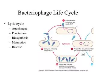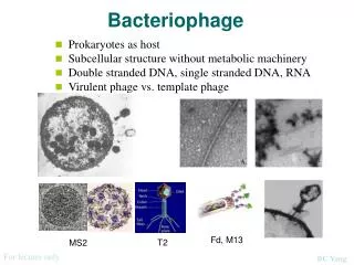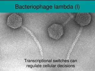
Bacteriophage
Bacteriophage. Maddy and Megan . Introduction. Our goal was to isolate, purify and characterize our phage from a specific environmental sample. We are isolating the phage to be able to study the different genetic sequences. It may be useful in the study of bacterial diseases


Bacteriophage
E N D
Presentation Transcript
Bacteriophage Maddy and Megan
Introduction • Our goal was to isolate, purify and characterize our phage from a specific environmental sample. • We are isolating the phage to be able to study the different genetic sequences. • It may be useful in the study of bacterial diseases • May also be useful with phage therapy
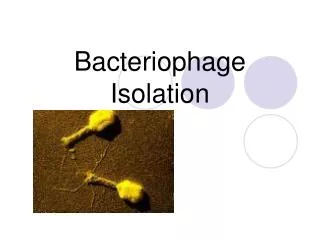
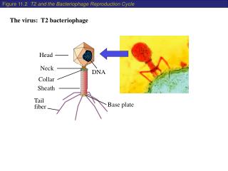
Methods • We obtained a soil sample for phage isolation • We then enriched the soil sample • Then we harvested and prepared the sample in order to obtain plaques • Next we processed plaques • Then determined phage titer and made streak plates • Then purified the phage by titrating the sample two more times • We then titrated our sample a fourth time in order to get web plates and determined phage titer • Then we harvested a phage lysate by flooding our web plates and then usuing that we did more titration and made more plates • Then determined phage titer once again • Then we made more web plates using our MTL
methods • We then flooding our web plates and filtered out liquid to make our HTL • Next we made plates to determine our phage titer • Then we used HTL to degrade bacterial DNA • Then we determined phage titer • Finally we isolated our phage genomic DNA • We then quantified our DNA using a spectrophotometer • Then we electrophoresed our DNA to take pictures • Then digested our phage genomic DNA with various enzymes and electrophoresed the samples to determine how the enzymes cut our DNA
Godzilla • I found my original phage about a foot away from the river near the amphitheater. • It was at 44.8594555 latitude, -92.6218815 longitude • in a moist, 65mm depth, at 77 degrees
Godzilla • I wasn’t able harvest any plaques from my soil sample so I then picked a plaque from one the Hudson phages • I originally found two different types, one was a small plaque and the other was a large one • During the process of purifying our phage, they both grew to be the same size • I then gave my other samples to Megan, because hers died
Godzilla • My first titer was 1.14×10^6 pfu/mL • My second titer was 1.09×10^7 pfu/mL • My third titer was 6.2×10^7 pfu/mL • My fourth titer was 4.96×10^9 pfu/mL • I my final titer it was very low .061 pfu/mL in each one of my titer calculations
Godzilla • My first phage genomic DNA electrophoresed sample was only cut by Hind3 • My second genomic DNA electrophoresed was cut by Nco1
Godzilla • I used the website to look at the phage Salgado, and found mine was not cut the same • No enzymes related to my phage
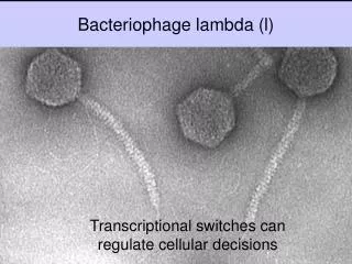
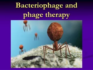
My phage measures about 25nm And the tail measures about 50nm
Toulouse • My soil sample did not yield plaques • Used Hudson phages and did a titration • First titer: 9.87×10⁴pfu/mL • Second titer: 1.494×10⁷pfu/mL • Third titer: 2.7×10⁴pfu/mL • Lost my phage during harvesting the phage lysate and titration • Adopted Maddy’s smaller phage
Pictures of spot test and my 10-4 and 10-5 plate My titer was 1.265×10⁹
Then electrophoresed our DNA • Quantified DNA using spectrophotometer • .2545 μg/μL
Digested our phage DNA and electrophoresed it My phage’s DNA was cut by Hind III and NcoI
Conclusion • We think that our phage might be the same. • We were not able to find any other plaques that were similar to ours however ours were similar