Unit 5 - Genetics
Unit 5 - Genetics. Topic 4 and 10 - Genetics. Topic 4 - genetics. 4.1 - Chromosomes, genes, alleles and mutations . 4.1.1 Eukaryotic Chromosomes 4.1.2 Definitions 4.1.3 Gene mutation. 4.1.4 Sickle cell anaemia. 4.1.1 State that eukaryotic chromosomes are made of DNA and protein.

Unit 5 - Genetics
E N D
Presentation Transcript
Unit 5 - Genetics Topic 4 and 10 - Genetics
4.1 - Chromosomes, genes, alleles and mutations • 4.1.1 Eukaryotic Chromosomes • 4.1.2 Definitions • 4.1.3 Gene mutation. • 4.1.4 Sickle cell anaemia
4.1.1 State that eukaryotic chromosomes are made of DNA and protein • Eukaryotic chromosomes consist of DNA wrapped around histone proteins • This forms the basic structure of the nucleosome, which is packed together to form chromatin (in a 'beads on a string' arrangement) • Chromatin will supercoil and condense during prophase to form chromosomes that can be visualised under a light microscope • Prokaryotic DNA is not wrapped around proteins and is thus considered to be 'naked'
4.1.2 Define gene, allele and genome • Gene: A heritable factor that controls a specific characteristic, consisting of a length of DNA occupying a particular position on a chromosome (locus) • Allele: One specific form of a gene, differing from other alleles by one or a few bases only and occupying the same locus as other alleles of the gene • Genome: The whole of the genetic information of an organism
4.1.3 Define gene mutation • Gene mutation: A change in the nucleotide sequence of a section of DNA coding for a particular feature
TYPES OF MUTATIONS • http://highered.mcgraw-hill.com/sites/0072556781/student_view0/chapter11/animation_quiz_4.html
4.1.4 Explain the consequence of a base substitution mutation in relation to the process of transcription and translation using the example of sickle cell anaemia • Cause of Sickle Cell Anemia • A base substitution mutation is the change of a single base in a sequence of DNA, resulting in a change to a single mRNA codon during transcription • In the case of sickle cell anemia, the 6th codon for the beta chain of hemoglobin is changed from GAG to GTG (on the non-coding strand) • This causes a change in the mRNA codon (GAG to GUG), resulting in a single amino acid change of glutamic acid to valine (Glu to Val) • DNA: GAG to GTG (non-coding strand) • mRNA: GAG to GUG • Amino Acid: Glu to Val • The amino acid change alters the structure of hemoglobin, causing it to form fibrous, insoluble strands • This causes the red blood cell to adopt a sickle shape
Consequences of Sickle Cell Anemia • The insoluble hemoglobin cannot effectively carry oxygen, causing individual to feel constantly tired • The sickle cells may accumulate in the capillaries and form clots, blocking blood supply to vital organs and causing a myriad of health problems • Also causes anemia (low RBC count), as the sickle cells are destroyed more rapidly than normal red blood cells • Sickle cell anemia occurs in individuals who have two copies of the codominant 'sickle cell' allele (i.e. homozygotes) • Heterozygous individuals have increased resistance to malaria due to the presence of a single 'sickle cell' allele (heterozygous advantage)
4.2 - Meiosis • 4.2.1 Reduction division • 4.2.2 Homologous Chromosomes • 4.2.3 Meiosis • 4.2.4 Non-disjunction • 4.2.5 Karyotyping • 4.2.6 Pre-natal diagnosis • 4.2.7 Human karyotyping.
4.2.1 State that meiosis is a reduction division of a diploid nucleus to form haploid nuclei Meiosis is the process by which sex cells (gametes) are made in the reproductive organs: • Most sexually reproducing animals are diploid - meaning they have two copies of every chromosome (one of maternal origin, one of paternal origin) • In order to reproduce, these organisms need to make gametes that are haploid (have only one copy of each chromosome) • Fertilization of two haploid gametes (egg + sperm) will result in the formation of a diploid zygote that will grow into a new organism Meiosis consists of two cell divisions: 1) The first division is a reduction division of the diploid nucleus to form haploid nuclei 2) The second division separates sister chromatids (this division is necessary because meiosis is preceded by interphase, wherein DNA is replicated)
4.2.2 Define homologous chromosomes • Homologous chromosomes are chromosomes that share: • The same structural features (e.g. same size, same banding pattern, same centromere position) • The same genes at the same loci positions (while genes are the same, alleles may be different)
4.2.3 Outline the process of meiosis, including pairing of homologous chromosomes and crossing over, followed by two divisions, which results in four haploid cells • The process of meiosis involves two divisions, both of which follow the same basic stages as mitosis (prophase, metaphase, anaphase and telophase) • Meiosis is preceded by interphase, which includes the replication of DNA (S phase) to create chromosomes with genetically identical sister chromatids • Meiosis I • Homologous chromosomes must first pair up in order to be sorted into separate haploid daughter cells • In prophase I, homologous chromosomes undergo a process called synapsis, whereby homologous chromosomes pair up to form a bivalent (or tetrad) • The homologous chromosomes are held together at points called chiasma (singular: chiasmata) • Crossing over of genetic material between non-sister chromatids can occur at these points, resulting in new gene combinations (recombination) • The remainder of meiosis I involves separating the homologous chromosomes into separate daughter cells • In metaphase I, the homologous pairs line up along the equator of the cell • In anaphase I, the homologous chromosomes split apart and move to opposite poles • In telophase I, the cell splits into two haploid daughter cells as cytokinesis happens concurrently
Meiosis II • In meiosis II, the sister chromatids are divided into separate cells • In prophase II, spindle fibers reform and reconnect to the chromosomes • In metaphase II, the chromosomes line up along the equator of the cell • In anaphase II, the sister chromatids split apart and move to opposite poles • In telophase II, the cell splits in two as cytokinesis happens concurrently • Because sister chromatids may no longer be genetically identical as a result of potential recombination, the process of meiosis results in the formation of four genetically distinct haploid daughter cells
Meiosis video • Animation - http://highered.mcgraw-hill.com/sites/0072495855/student_view0/chapter28/animation__how_meiosis_works.html • http://www.youtube.com/watch?v=qCLmR9-YY7o – 11 min
4.2.4 Explain that non-disjunction can lead to a change in chromosome number, illustrated by reference to Down syndrome (trisomy 21) • Non-disjunction refers to the chromosomes failing to separate correctly, resulting in gametes with one extra, or one missing, chromosome (aneuploidy) • The failure of the chromosomes to separate may either occur via: • Failure of homologues to separate during Anaphase I (resulting in four affected daughter cells) • Failure of sister chromatids to separate during Anaphase II (resulting in two affected daughter cells)
Individuals with Down syndrome have three copies of chromosome 21 (trisomy 21) • One of the parental gametes had two copies of chromosome 21 as a result of non-disjunction • The other parental gamete was normal and had a single copy of chromosome 21 • When the two gametes fused during fertilization, the resulting zygote had three copies of chromosome 21, leading to Down syndrome
4.2.5 State that, in karyotyping, chromosomes are arranged in pairs according to their structure • karyotype is a visual profile of all the chromosomes in a cell • The chromosomes are arranged into homologous pairs and displayed according to their structural characteristics • Karyotyping involves: • Harvesting cells (usually from fetus or white blood cells of adults) • Chemically inducing cell division, then halting it during mitosis when chromosomes are condensed and thus visible • The stage during which mitosis is halted will determine whether chromosomes appear with sister chromatids • Staining and photographing chromosomes, before arranging them according to structure
4.2.6 State that karyotyping is performed using cells collected by chorionic villus sampling or amniocentesis, for pre-natal diagnosis of chromosome abnormalities • Pre-natal karyotyping is often used to: • Determine the gender of an unborn child (via identification of sex chromosomes) • Test for chromosomal abnormalities (e.g. aneuploidies resulting from non-disjunction) • Amniocentesis • A needle is inserted through the abdominal wall, into the amniotic cavity in the uterus, and a sample of amniotic fluid containing fetal cells is taken • It can be done at ~ 16th week of pregnancy, with a slight chance of miscarriage (~0.5%) • Chorionic Villus Sampling • A tube is inserted through the cervix and a tiny sample of the chorionic villi (contains fetal cells) from the placenta is taken • It can be done at ~ 11th week of pregnancy, with a slight risk of inducing miscarriage (~1%)
Amniocentesis Chorionic Villus Sampling http://www.youtube.com/watch?v=bZcGpjyOXt0 http://video.about.com/pregnancy/Chorionic-Villus-Sampling.htm http://www.youtube.com/watch?v=xwZOmVsiOHI
4.2.7 Analyze a human karyotype to determine gender and whether non-disjunction has occurred • Every cell in the human body has 46 chromosomes (except anucleate red blood cells and haploid gametes) • Males (X,Y) and females (X,X) can be differentiated on the basis of their sex chromosomes • Non-disjunction during gamete formation can lead to individuals with an abnormal number of chromosomes (aneuploidy) • These disorders can be classified according to the chromosome number affected and the number of chromosomes present
4.3 – Theoretical Genetics • 4.3.1 Definitions • 4.3.2 Monohybrid crosses • 4.3.3 Multiple alleles. • 4.3.4 Codominance & Multiple alleles (ABO blood groups). • 4.3.5 Sex chromosomes. • 4.3.6 Sex chromosomes and genes. • 4.3.7 Sex Linkage. • 4.3.8 Colourblindness and haemophilia. • 4.3.9 Female and sex linkage. • 4.3.10 Females & X-linked recessive alleles. • 4.3.11 Predicting genotypic and phenotypic ratios. • 4.3.12 Pedigree
4.3.1 Define genotype, phenotype, dominant allele, recessive allele, codominant alleles, locus, homozygous, heterozygous, carrier and test cross • Genotype: The allele combination of an organism • Phenotype: The characteristics of an organism (determined by a combination of genotype and environmental factors) • Dominant Allele: An allele that has the same effect on the phenotype whether it is present in the homozygous or heterozygous state • Recessive Allele: An allele that only has an effect on the phenotype when present in the homozygous state • Codominant Alleles: Pairs of alleles that both affect the phenotype when present in a heterozygote • Locus: The particular position on homologous chromosomes of a gene • Homozygous: Having two identical alleles of a gene • Heterozygous: Having two different alleles of a gene • Carrier: An individual that has one copy of a recessive allele that causes a genetic disease in individuals that are homozygous for this allele • Test Cross: Testing a suspected heterozygote by crossing it with a known homozygous recessive
4.3.2 Determine the genotypes and phenotypes of the offspring of a monohybrid cross using a Punnett grid • A genetic cross is a means of determining the genetic characteristics of potential offspring based on the genetic characteristics of the prospective parents • A monohybrid cross determines the allele combinations of offspring for one particular gene only (HL students may refer to topic 10.2 for dihybrid crosses) • Monohybrid crosses can be calculated according to the following steps: • Step 1: Designate characters to represent the alleles • Capital letter for dominant allele, lower case letter for recessive allele • Step 2: Write down the genotype and phenotype of the parents • This is the P generation (parental generation) • Step 3: Write down the genotype of the parental gametes • These will be haploid as a result of meiotic division • Step 4: Use a Punnett grid to work out the potential gamete combinations • As fertilization is random, all combinations have an equal probability • Step 5: Write out the genotype and phenotype ratios of potential offspring • This is the F1 generation (first filial generation) • Subsequent generations through interbreeding labeled F2, F3, etc.
4.3.3 State that some genes have more than two alleles (multiple alleles) • Some genes have more than two alleles for a given trait (e.g. the ABO blood group system) • The alleles which are not recessive may either: • Share codominance (be expressed equally in the phenotype) • Share incomplete dominance (neither is fully expressed in the phenotype, resulting in blending) • Demonstrate a dominance order (e.g. allele A > allele B > allele C)
4.3.4 Describe ABO blood groups as an example of codominance and multiple alleles • When assigning alleles for codominance, the convention is to use a common letter to represent dominant and recessive and use superscripts to represent the different codominant alleles • I stands for immunoglobulin (antigenic protein on blood cells) • A and B stand for the codominant variants • The ABO gene has three alleles: IA, IB and i • IA and IB are codominant, whereas i is recessive (no antigenic protein is produced) • Codominance means that both IA and IB alleles will be expressed within a given phenotype
4.3.5 Explain how sex chromosomes control gender by referring to the inheritance of X and Y chromosomes in humans • Humans have 23 pairs of chromosomes for a total of 46 (excluding instances of aneuploidy) • The first 22 pairs are autosomes - each chromosome pair possesses the same genes and structural features • The 23rd pair of chromosomes are heterosomes (or sex chromosomes) and determine gender • Females are XX - they possess two X chromosomes • Males are XY - they posses one X chromosome and a much shorter Y chromosome • The Y chromosome contains the genes for developing male sex characteristic - hence the father is always responsible for determining gender • If the male sperm contains the X chromosome the growing embryo will develop into a girl • If the male sperm contains a Y chromosome the growing embryo will develop into a boy • In all cases the female egg will contain an X chromosome (as the mother is XX) • Because the X and Y chromosomes are of a different size, they cannot undergo crossing over / recombination during meiosis • This ensures that the gene responsible for gender always remains on the Y chromosome, meaning that there is always ~ 50% chance of a boy or girl
4.3.6 State that some genes are present on the X chromosome and absent from the shorter Y chromosome • The Y chromosome is much shorter than the X chromosome and contains only a few genes • Includes the SRY sex-determination gene and a few others (e.g. hairy ears gene) • The X chromosome is much longer and contains several genes not present on the Y chromosome • Includes the genes for hemophilia and red-green color blindness • In human females, only one of the X chromosomes remains active throughout life • The other is packaged as heterochromatin to form a condensed Barr body • This inactivation is random and individual to each cell, so heterozygous women will be a mosaic - expressing both alleles via different cells
4.3.7 Define sex linkage • Sex linkage refers to when a gene controlling a characteristic is found on a sex chromosome (and so we associate the trait with a predominant gender) • Sex-linked conditions are usually X-linked, as very few genes exist on the shorter Y chromosome
4.3.8 Describe the inheritance of color blindness and hemophilia as examples of sex linkage • Color blindness and hemophilia are both examples of X-linked recessive conditions • The gene loci for these conditions are found on the non-homologous region of the X chromosome (they are not present of the Y chromosome) • As males only have one allele for this gene they cannot be a carrier for the condition • This means they have a higher frequency of being recessive and expressing the trait • Males will always inherit an X-linked recessive condition from their mother • Females will only inherit an X-linked recessive condition if they receive a recessive allele from both parents • When assigning alleles for sex-linked traits the convention is to write the allele as a superscript to the sex chromosome (usually X) • Hemophilia: XH = unaffected ; Xh = affected • Color Blindness: XA = unaffected ; Xa = affected
4.3.9 State that a human female can be homozygous or heterozygous with respect to sex-linked genes • As human females have two X chromosomes (and therefore two alleles for any given X-linked gene), they can be either homozygous or heterozygous • Males only have one X chromosome (and therefore only one allele) and are hemizygous
4.3.10 Explain that female carriers are heterozygous for X-linked recessive alleles • An individual with a recessive allele for a disease condition that is masked by a normal dominant allele is said to be a carrier • Carriers are heterozygous and can potentially pass the trait on to the next generation, but do not suffer from the defective condition themselves • Females can be carriers for X-linked recessive conditions because they have two X chromosomes - males (XY) cannot be carriers • Because a male only inherits an X chromosome from his mother, his chances of inheriting the disease condition from a carrier mother is greater
4.3.11 Predict the genotypic and phenotypic ratios of offspring of monohybrid crosses involving any of the above patterns of inheritance Autosomal Dominance / Recessive • Choose a letter where the upper and lower case forms are easily distinguishable (e.g. E/e, A/a, B/b) • Use the capital letter for the dominant allele and the lower case letter for the recessive allele • Example:
Codominance • Choose a letter to denote the general trait encoded by the gene (capital = dominant, lower case = recessive) • Use different superscript letters (capitals) to represent the different codominant alleles • Example:
X-linked Recessive • Use a capital "X" to denote the X chromosome • Choose a superscript letter to represent the trait (capital = dominant, lower case = recessive) • Example:
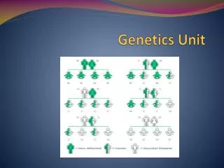
4.3.12 Deduce the genotype and phenotype of individuals in pedigree charts • A pedigree is a chart of the genetic history of a family over several generations • Males are represented as squares, while females are represented as circles • Shaded symbols means an individual is affected by a condition, while an unshaded symbol means they are unaffected • A horizontal line between a man and woman represents mating and resulting children are shown as offshoots to this line
Autosomal Dominance • All affected individuals must have at least one affected parent • If two parents are unaffected, all offspring must be unaffected (homozygous recessive) • If two parents are affected, they may have offspring who are unaffected (if parents are heterozygous)
Autosomal Recessive • If two parents show a trait, all children must also show the trait (homozygous recessive) • An affected individual may have two normal parents (if parents are both heterozygous carriers)
X-Linked Recessive • If a female shows the trait, so must all sons as well as her father • The disorder is more common in males
4.4 – Genetic Engineering and Biotechnology • 4.4.1 Polymerase chain reaction • 4.4.2 Gel electrophoresis • 4.4.3 DNA profiling • 4.4.4 DNA profiling and applications in paternity and forensic investigations. • 4.4.5 Interpretation of paternity and forensic investigations. • 4.4.6 Human genome project. • 4.4.7 Gene transfer • 4.4.8 Gene transfer techniques. • 4.4.9 Genetically modified crops and animals • 4.4.10 Hazards and benefits of genetic modification. • 4.4.11 Definition of clone. • 4.4.12 Cloning in differentiated animal cells. • 4.4.13 Ethical issues of therapeutic cloning