General Genetics
General Genetics. Ayesha M. Khan Spring 2013. X-linked color blindness in humans Light detecting cells: Rods – See in shades of grey Cones – See in colors

General Genetics
E N D
Presentation Transcript
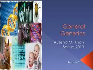
General Genetics Ayesha M. Khan Spring 2013
X-linked color blindness in humans • Light detecting cells: • Rods – See in shades of grey • Cones – See in colors • Within the human eye, color is perceived in light sensing cone cells that line the retina. Each cone cell contains one of three pigments capable of absorbing light of a particular wavelength; one absorbs blue light, a second absorbs red light, and a third absorbs green light. X-Linked Characteristics
X-linked color blindness in humans What a red-green color-blind person sees. What people with normal color vision see. How Color-Blind People See Things
The human eye actually detects only three colors—red, green, and blue—but the brain mixes the signals from different cone cells to create the wide spectrum of colors that we perceive. • Each of the three pigments is encoded by a separate locus; the locus for the blue pigment is found on chromosome 7, and those for green and red pigments lie close together on the X chromosome. • Mutations that produce defective color vision are generally recessive and, because the genes coding for the red and green pigments are located on the X chromosome, red–green color blindness is inherited as an X-linked recessive characteristic. X-linked color blindness in humans
Crisscross inheritance • Pattern of inheritance exhibited by X-linked recessive characteristics • Appearing in females one generation and in males the next generation
a. Affected fathers transmit the recessive allele to all daughters (who are therefore carriers), and to none of their sons. b. Father-to-son transmission of X-linked alleles generally does not occur. c. Many more males than females exhibit the trait. d. All sons of affected (homozygous recessive) mothers are expected to show the trait. e. With a carrier mother, about 1⁄2 of her sons will show the trait and 1⁄2 will be free of the allele. f. A carrier female crossed with a normal male will have 1⁄2 carrier and 1⁄2 normal daughters. Some characteristics of X-linked recessive inheritance:
Drosophila with white eyes: Female’s genotype-> ww OR XwXw Male’s genotype-> w/ OR XwY Symbols for X-Linked Genes
Hemophilia (reduced blood clotting) is an X-linked recessive disease in humans. A woman with hemophilia mates with a man who exhibits normal blood clotting. 1) What is the probability that their child will have hemophilia? 2) What is the probability that their male child will have hemophilia? Concept check
Different numbers of X chromosomes in males and females -potential problem. • The amount of protein produced by X-linked genes would differ in the two sexes. • Females would produce twice as much • This difference could be highly detrimental Equalizes the amount of protein produced by X-linked genes in the two sexes. Dosage Compensation
As a result of X inactivation, females are functionally hemizygousat the cellular level for X-linked genes. • In females that are heterozygous at an X-linked locus, approximately 50% of the cells will express one allele and 50% will express the other allele; thus, in heterozygous females, proteins encoded by both alleles are produced, although not within the same cell Barr body Murray Barr (1949) Observed condensed, darkly staining bodies in the nuclei of cells from female cats. Dosage Compensation
An interphase epithelial cell from a human female shows a dark-staining Barr body in the nucleus; representing an inactivatedX chromosome.A comparable cheek epithelial cell from a male does not show a Barr body. Thus, dosage compensation prevents excessive expression of X-linked genes in mammals.
Lyon hypothesis Mary Lyon (1961) Within each female cell, one of the two X chromosomes becomes inactive; which X chromosome is inactivated is random. If a cell contains more than two X chromosomes, all but one of them is inactivated.
Number of Barr bodies in human cells with different complements of sex chromosomes
In heterozygous females, proteins encoded by both alleles are produced, although not within the same cell. • Females are functionally hemizygousat the cellular level for X-linked genes. • Females are mosaics for the expression of X-linked genes. Result of X inactivation:
Black and orange patches Example: Tortoiseshell cats
A single X-linked locus determines the presence of orange color. • X+ which produces nonorange (usually black) fur • Xo which produces orange fur Males are hemizygous and thus may be black (X+ Y) or orange (Xo Y) but not black and orange. Females may be black (X+ X+ ), orange (XoXo), or tortoiseshell (X+ Xo ). Example: Tortoiseshell cats
Inheritance of Z-linked characteristics is the same as that of X-linked characteristics, except that the pattern of inheritance in males and females is reversed. -Females are heterogametic sex Z-Linked Characteristics