Download
1 / 21
260 likes | 793 Vues
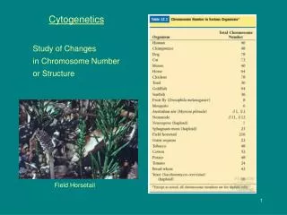
Cytogenetics General Genetics. Dr. Attya Bhatti. Cytogenetics. Is the study of the structure and properties of chromosomes , chromosomal behaviour during mitosis and meiosis , chromosomal influence on the phenotype and the factors that cause chromosomal changes.

E N D
CytogeneticsGeneral Genetics Dr. Attya Bhatti
Cytogenetics • Is the study of the structure and properties of chromosomes, chromosomal behaviour during mitosis and meiosis, chromosomal influence on the phenotype and the factors that cause chromosomal changes. • Related to disease status caused by abnormal chromosome number and/or structure.
Methods for chromosomal analysis: Karyotyping and banding The collection of all the chromosomes is referred to as a Karyotype. The method used to analyze the chromosome constitution of an individual, known as chromosome banding. Chromosomes are displayed as a karyogram.
Obtaining and preparing cells for chromosome analysis • Cell source: • Blood cells • Skin fibroblasts • Amniotic cells / chorionic villi • Increasing the mitotic index - proportion of cells in mitosis using colcemid • Synchronizing cells to analyze prometaphase chromosomes
Key procedure • In the case of peripheral (venous) blood • A sample is added to a small volume of nutrient medium containing phytoheamagglutinin, which stimulates T lymphocytes to divide. • The cells are cultured under sterile conditions at 37C for about 3 days, during which they divide, and colchicineis then added to each culture. • This drug has the extremely useful property of preventing formation of the spindle, thereby arresting cell division during metaphase, the time when the chromosomes are maximally condensed and therefore most visible. • Hypotonic saline is then added, which causes the red blood cells to lyze and results in spreading of the chromosomes, which are then fixed , mounted on a slide and stained ready for analysis
Karyotype Analysis • Following Steps are involved; • Counting the number of cells, sometimes referred as metaphase spread • Analysis of the banding pattern of each individual chromosome in selected cells. • Total chr. Count is determined in 10-15 cells, but if mosaicism is suspected then 30 or more cell count will be undertaken. • Detailed analysis of the banding pattern of the individual chromosomes is carried out in approx. 3-5 metaphase spread, which shows high quality banding. • The banding pattern of each chromosome is specific and shown in the form of Idiogram.
Chromosome Banding • Chromosome banding is developed based on the presence of heterochromatin and euchromatin. • Heterochromatin is darkly stained whereas euchromatin is lightly stained during chromosome staining. • Euchromatin, which undergoes the normal process of condensation and decondensation in the cell cycle, and • Heterochromatin, which remains in a highly condensed state throughout the cell cycle, even during interphase.
Types of chromosome banding • G-banding • C-banding • Q-banding • R-banding • T-banding
Chromosomal Banding • G-banding, gives dark bands • C-banding: C-banding stains the constitutive heterochromatin, which usually lies near the centromere. • Q-banding: Q-banding is a fluorescent pattern obtained using quinacrine for staining. The pattern of bands is very similar to that seen in G-banding. • R-banding: reverse of G-banding (the R stands for "reverse"). • Dark regions are euchromatic (guanine-cytosine rich regions) and the bright regions are heterochromatic (thymine-adenine rich regions). • T-banding:Identifies a subset of the R bands which are especially concentrated at the telomeres.
G-Banding • - G-banding is obtained with Giemsa stain following digestion of • chromosomes with enzyme trypsin. • - Giemsa stain, named after Gustav Giemsa, an early malariologist, is • used for the histopathological diagnosis of malaria and other parasites. • - It is a mixture of methylene blue and eosin. • - It is specific for the phosphate groups of DNA and attaches itself to • regions of DNA where there are high amounts of adenine-thymine • bonding. • - it yields a series of lightly and darkly stained bands – the dark regions • tend to be heterochromatic, late-replicating and AT rich. • - The light regions tend to be euchromatic, early-replicating • and GC rich.
Key procedure • In the case of peripheral (venous) blood • A sample is added to a small volume of nutrient medium containing phytoheamagglutinin, which stimulates T lymphocytes to divide. • The cells are cultured under sterile conditions at 37C for about 3 days, during which they divide, and colchicineis then added to each culture. • This drug has the extremely useful property of preventing formation of the spindle, thereby arresting cell division during metaphase, the time when the chromosomes are maximally condensed and therefore most visible. • Hypotonic saline is then added, which causes the red blood cells to lyze and results in spreading of the chromosomes, which are then fixed , mounted on a slide and stained ready for analysis
Molecular Methods for chromosomal analysis Molecular Cytogenetics • Fluorescent in situ Hybridization (FISH) • Chromosome painting • Comparative Genomic Hybridization (CGH) • Molecular karyotyping and Multiplex FISH(M-FISH) • Spectral Karyotyping • Array CGH
Key points • Cytogenetic analysis usually focuses on chromosomes in dividing cells. • Dyes such as Quinacrine and Giemsa create banding patterns that’s are useful in identifying individual chromosomes within a cell. • A karyotype shows the photographed chromosomes of a cell arranged for cytogenetic analysis.