Download
1 / 43
470 likes | 1.03k Vues
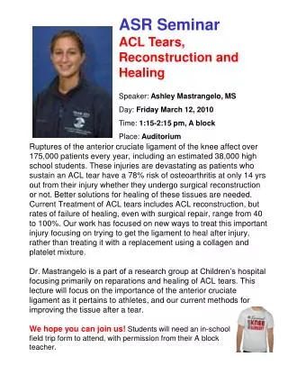
ACL Tears and Osteoarthritis. Emily Jones, MD Grand Rounds June 11, 2009. Objectives. What causes Osteoarthritis after ACL tears? Role of the Articular Cartilage Role of the Meniscus Does surgical intervention affect the progression of Osteoarthritis?. Our patient…. 40 y.o. male

E N D
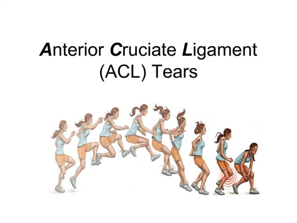
ACL Tears andOsteoarthritis Emily Jones, MD Grand Rounds June 11, 2009
Objectives • What causes Osteoarthritis after ACL tears? • Role of the Articular Cartilage • Role of the Meniscus • Does surgical intervention affect the progression of Osteoarthritis?
Our patient…. • 40 y.o. male • Worsening lateral left knee pain when he plays soccer • No acute injury • Hx of ACL tear at age 29 • ACL reconstructed with BTB graft • Not sure if he had any other damage at the time of initial injury 11 years ago
Physical Exam • Fit, BMI 23 • Normal gait • No swelling, erythema • Tenderness lateral joint line • ROM 0 to 125 • Equivocal Steinmann, McMurray • Lachman 1+ bilaterally • Negative valgus/varus
Differential Diagnosis • Repeat ACL tear • Meniscal Tear • Osteoarthritis • Contusion • LCL sprain • IT band
Probable Diagnosis… Osteoarthritis
Epidemiology • 1 in 3,500 U.S. residents tear ACL each year = 95,000/year • Development of osteoarthritis after ACL tear • 45% within 10 years (42,750 develop knee OA within 10 yrs) • 60 to 90% within 10 to 15 years • 16 to 90% in 10 to 15 years • Onset 10 to 20 years earlier in non-ACL knee • Estimated that acl rupture ages knee 30 years • ACL rupture >5 fold increase risk of knee OA • RR of 1.7 for increased weight
Epidemiology OA Soccer • 67 Female soccer players • 12 yrs after ACL tear • Average age 31, BMI 23 • 60% had reconstruction • 82 % radiographic changes in knee • 51% had changes consistent with OA • 75% sxs affecting their quality of life • Surgical reconstruction no effect
Epidemiology OA Soccer • Male soccer players s/p ACL tear • 219 patients, 14 years later • Radiographic changes 78% injured knees • Kellgren-Lawrence grade 2 or higher41% • seen in 4% of uninjured knees • 80% had reduced activity level and said knees contributed to this • No difference with surgical reconstruction
Iatrogenic ACL tear Model • ACL-transection (ACLT) model is extensively used to study osteoarthritis and its treatment • Ligament is sectioned to initiate osteoarthritis • Biochemical changes to the cartilage shown to occur within 3 weeks of the surgical intervention • Bone marrow edema beneath the medial compartment of the tibial plateau is detected with MRI within 6 weeks of ACLT in the dog • Evidence of cartilage erosion by the 12th week • Osteophytosis and subsequent meniscal damage occur by the 24th week • Long-term progression has been shown to mimic that of humans
Acute injury Risk • ACL tear is usually a non-impact, rotational injury • Tibia subluxes anteriorly • Impacts the lateral femoral condyle • High shear forces across tibiofemoral articular cartilage
Articular Cartilage • Extracellular matrix surrounding chondrocytes • Chondrocytes are responsible for production and maintenance of extracellular matrix • ECM primarily type 2 cartilage • 90-95% of the collagen present in articular cartilage • Designed to help resist shear stress • Articular cartilage continually undergoes metabolic activity to synthesize + degrade its matrix • Disrupted by injury • Cartilage is aneural and avascular • Chondrocyte viability depends on diffusion of metabolites from synovial fluid
Articular Cartialge • Articular cartilage is 4 layers: superficial, middle, deep, calcified • Tidemark = calcified cartilage separated from non-calcified deep cartilage layer • Cadaver models have shown that tidemark may be the weak link to shear forces
Articular Cartilage • Likely to have insult to articular cartilage at time of ACL disruption • May not be visible initially • Especially if at tide mark • May progress to communicate with the joint surface and surface lesion may become evident • Study of ACL deficient knees and cartilage lesions • 40% at 1 year • 60% at 5 years • >80% at 10 years
Articular Cartilage • Injured cartilage does not heal • Chondrocytes loose ability to migrate • Acutely injured chondrocytes produce collagen and proteoglycans • Insufficient to fill injured region and does not result in hyaline cartilage • Fibrocartilage
Articular Cartilage • Gold standard for identifying chondral lesions is arthroscopic viewing and probing of articular surfaces • Visible lesion often only ~ 1/3 size total • Closed lesions – softened, +/- surface changes • Open – classified by size and depth • Full-thickness have exposed subchondral bone
How diagnose articular cartilage injuries? • Often not initially visible on initial arthroscopy (as previously stated) • MRI sensitivity ~ 21% • 3D SPGR up to 62% • What about a correlation with MRI finding of bone bruises?
MRI • Bone bruising seen on MRI ~ 80% of the time with ACL tear • Location: posteriorlateral tibia + lateral femoral condyle • Reticular lesion in >70% = less severe hemm and edema, minimal damage to subchondral bone • geographic lesions ~25% = more severe – changes in subchondral bone • Correlated with histological degeneration of overlying chondrocytes and loss of proteoglycan component of the cartilage
MRI • 3D MRSI and T1 mapping • Difference in bone marrow bruising lateral tibia s/p ACL seen • Arthroscopic softening posteriorlateral tibia corresponding
The Studies Bone Bruising + Articular Cartilage • Study: Articular Cartilage Injury of the Posterior Lateral Tibial Plateau associated with acute ACL Injury • 39 patients with recent ACL rupture • Findings - statistically significant correlation b/w proportion of bone bruise and cartilage injury of the lateral femoral condyle,the posterior lateral tibial plateau and that of tears in the LM posterior horn seen on arthroscopy during ACL reconstruction • Conclusion: Pay attention to cartilage damage of the posterior lateral tibial plateau, lateral femoral condyle, as well as to posteriorhorn tears in LM with acute ACL when bone bruising seen on MRI
Causes of Osteoarthritis after ACL tear • #1 Acute Articular Chondral damage • No evidence that treating these changes the progression of osteoarthritis • Possible role for fixing ACL to decrease shearing forces overtime. More on this later……
What about the meniscus? • Meniscal pathology resulting in menisectomy is known to correlate with joint degeneration • Menisectomy might be the most important risk factor for developing TF OA s/p ACL • Not known if menisectomy risk for PF OA • Stable reconstructed knees with intact menisci also progress to OA more rapidly
The Meniscal/ACL studies • ACL Reconstruction After 10 to 15 Years, Association between Meniscectomy and Osteoarthritis • Reconstruction used patellar graft • Statistically significant association between medial or lateral OA and meniscal injury • If had meniscal injury at time of reconstruction – developed OA in the compartment where the meniscal injury was by 10 to 15 years out • Meniscectomy was also assoc with poorer results on objective test of knee fx even with stable joint
The Meniscal/ACL studies • Study:Primary ACL reconstruction vs no surgical treatment for ACL • If menisectomy was performed – 2/3 showed OA changes (vs 15% overall in study) regardless of initial treatment of ACL with surgical or non-surgical • This included PF OA in addition to TF OA
The Meniscal/ACL studies • Study: Starting with OA and seeing if lack of ACL incidentally matters in progression • 265 patients with symptomatic OA over 30-months • Presence of complete ACL tear at baseline increased risk for greater cartilage loss at medial tibiofemoral compartment • But, once presence of concurrent medial meniscal tears was taken into account, no independent risk of complete ACL tear on cartilage loss • Trend for an increase in cartilage loss at lateral compartment – also related to meniscal pathology in lateral compartment • No assoc b/w complete ACL tear and cartilage loss at patellofemoral joint • Meniscal tears more frequent in those without ACL – not known if happened at time of ACL or afterwards
The Meniscal/ACL studies • Conclusion: findings suggest that concurrent meniscal pathology, which may have occurred at the time of possible knee injury causing the ACL tear, or which resulted from an ACL tear, or which was independent of the ACL tear, is responsible for the accelerated cartilage loss, at least seen in short-term follow-up.” • How such individuals – with symptomatic knee OA, acl tear, accelerated cartilage loss at medial tibiofemoral compartment due to concomitant meniscal tears – should be tx’d not determined.
Causes of Osteoarthritis after ACL tear • #1 Acute Articular Chondral damage • No evidence that treating these changes the progression of osteoarthritis • Possible role for fixing ACL to decrease shearing forces overtime. More on this later…… • #2 Acute Meniscal Injury • Early ACL reconstruction can reduce risk of secondary meniscal tears
Surgical reconstruction • Does improve stability • Probably decreases new meniscal and chondral injuries • Not proven to prevent long term OA • Patellar Tendon Graft = increase harvest-site symptoms and PF radiographic osteoarthritis vs hamstring tendon graft
The Surgical Reconstruction Studies • Function, OA and activity after ISOLATED ACL-rupture: 11 years follow-up results of conservative versus reconstructive tx • 109 patients – 60 reconstructions (bone-tendon-bone graft) • Retrospective cohort • Similar physical activity • ACL reconstruction = increased stability, increased OA (42 vs 25%) • Decreased secondary meniscal tears in reconstructed ACLs
The Surgical Reconstruction Studies Study: Long-term results after primary repair or non-surgical treatment of anterior cruciate ligament rupture: a randomized study with a 15-year follow-up • 100 patients, 15 yrs after random allocation to surgical repair vs. conservative • No difference activity level, knee injury, OA (subjective or radiographic) • More instability in non-surgical • One-third of the patients in the non-surgically treated group underwent secondary ACL reconstruction due to instability problems • This group had the most OA and secondary mensical injuries • Surgery does improve stability • Surgery did not decrease late OA (~50% regardless of treatment) • There were significantly more secondary meniscus injuries in patients initially treated non-surgically (12 vs 35%)
The Surgical Reconstruction Studies • Johma et al. reported that 11% of the patients who underwent acute ACL reconstruction and 50% of the patients who underwent chronic ACL reconstruction presented radiographic evidence of osteoarthritis after 7 years • suggests that cartilage damage becomes more severe as the time between injury and surgery increases • Daniel et al. in a 5-year prospective follow-up, determined that both acute and chronic ACL-reconstructed knees had significantly greater radiographic evidence of osteoarthritis compared with the conservatively treated group
The Surgical Reconstruction Studies • 100 patients with an acute ACL injury without reconstruction for observed for 15 years • concluded that early modification of activity and neuromuscular rehabilitation resulted in good knee function and an acceptable activity level in the majority of patients On the other hand…. • Strehl and Eggli, found that almost two-thirds of those patients initially selected for conservative treatment required surgical reconstruction in the long-term
The Surgical Reconstruction Studies • Study: Hamstring vs Patellar grafting for ACL reconstruction • Prospective Cohort • 90 hamstring grafts, 90 patellar grafts • At 10 years no significant difference in graft rupture rates (#7 vs #12) • In patellar group harvest site symptoms and kneeling pain more common, and more reported pain with strenuous activity • Radiographic OA more common in patellar at 10 years(P=0.04)
The Surgical Reconstruction Studies • Study: The effects of functional Knee brace during early tx of patients with a nonoperated acute ACL • Prospective randomized, 95 patients • 53 excluded (or dropped out) • Articular cartilage injury • Other injuries that negatively affected rehab • Desire for surgery • Patients experienced effect of the brace regarding sense of instability and rehab • No objective findings on knee OA outcome score, strength
Surgical Technique • Tunnel placement • Anterior tibial tunnel placement results in graft impingement against the intercondylar roof posterior tunnel= vertical graft = laxity • Anterior femoral tunnel places the graft under high tissue strains with knee flexion, resulting in decreased knee flexion or increased graft stretching • Inversely, the over-the-top position may cause the graft to tighten in the last degrees of extension and results in an increased anteroposterior laxity when knee is in flexion
Surgical technique • Notchplasty • Recent study showed that aggressive intercondylar notchplasty can cause articular cartilage histopathologic changes at 6 months, consistent with those found in knees with early degenerative arthrosis • A prospective, randomized study involving 100 patients found no beneficial short-term effect of performing a notchplasty - minimizing the notchplasty reduced the postoperative bleeding, pain, swelling, and potential notch regrowth • Therefore, extensive notchplasty or roofplasty should be performed only if deemed necessary after having tested the graft clearance intraoperatively
Causes of Osteoarthritis after ACL tear • #1 Acute Articular Chondral damage • No evidence that treating these changes the progression of osteoarthritis • Possible role for fixing ACL to decrease shearing forces overtime. More on this later…… • #2 Acute Meniscal Injury • Early ACL reconstruction can reduce risk of secondary meniscal tears • #3 Recurrent Instability • Improved by surgical reconstruction • ?Surgical Technique
Other factors – gait? • Study: Gait Mechanics in patients who underwent ACL reconstruction exhibited altered gait that may be associated with progression to OA • 17 patients • Dynamic frontal plane knee malalignment • Possibly promotes degredation of medial tibiofemoral compartment • Every 1% increase in internal abduction moment, risk of knee OA increases 6.46 times
Other Factors – gait? • Using stereofluoroscopy, Tashman et al. found that patients with reconstructed knees consistently run with their reconstructed knee externally rotated (by 3.8°) and more adducted (by 2.8°) than the control knee after 12 months of healing • Can’t reproduce original anatomy and kinematics with reconstruction
Due to lack of native ACL Technique? Type? At time Of injury
Summary • Osteoarthritis after ACL tear is prevalent • Articular cartilage • Likely to be damaged at time of injury • Likely to contribute to osteoarthritis • Meniscal injuries are one of the main determinants of developing OA • Surgery seems to decrease secondary meniscal injuries • Surgery improves stability • Reconstructive surgery has not been shown to decrease Osteoarthritis