Download
1 / 17
170 likes | 1.06k Vues
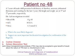
Patient no 24. A 24 years old female underwent abortion but she developed certain complications and was shifted to intensive care unit. Her laboratory investigations revealed:- ALT: 96 U/L PT: Normal: 14 sec Patient: 40 sec

E N D
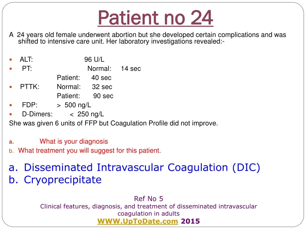
Patient no 24 A 24 years old female underwent abortion but she developed certain complications and was shifted to intensive care unit. Her laboratory investigations revealed:- • ALT: 96 U/L • PT: Normal: 14 sec Patient: 40 sec • PTTK: Normal: 32 sec Patient: 90 sec • FDP: > 500 ng/L • D-Dimers: < 250 ng/L She was given 6 units of FFP but Coagulation Profile did not improve. • What is your diagnosis • What treatment you will suggest for this patient. • Disseminated Intravascular Coagulation (DIC) • Cryoprecipitate Ref No 5 Clinical features, diagnosis, and treatment of disseminated intravascular coagulation in adults WWW.UpToDate.com 2015
Disseminated Intravascular Coagulation (DIC) • DIC is seen in approximately 1 percent of admissions to tertiary care hospitals. • DIC is a multi-organ disease requiring expertise of multi-specialty team • Chemical Pathologist often has to be the part of this team managing DIC.
Pathogenesis of DIC • The hallmark of DIC is coagulation and fibrinolysis occurring side by side but massively. • The process of coagulation overtakes the clotting factors synthesis process leading to so called ‘consumption coagulopathy’. • Massive fibrinolysis leads to formation of fibrin which form an important impediment in clotting.
Pathogenesis of DIC (cont) • Fibrin strands also cause fragmentation of RBC (Shistocytes & helmet cells) by ‘guilloting’ the RBCs • The term ‘chronic DIC’ is used for mild DIC without clotting defect and thrombocytopaenia.
Causes of DIC There are two basic causes of an episode of DIC • Release of tissue thromboplastin • Obstetric complications • AML – M3 • Intravascular haemolysis of various origins • Trauma, head injury, major surgery • Malignant tumours • Endothelial damage • Infections : Bacterial, viral, rickettsial, fungal, protozoal • Shock • Heat stroke • Snake venous • Drugs or antigen-antibody complexes
Lab Diagnosis of DIC • Blood CBC • RBC morphology • Abnormal cells/blasts • Leucoerythroblastic picture • Thrombocytopenia • PT, PTTK, TT prolonged in acute syndrome • Factor V and VIII are reduced • Fibrinogen level is low • High levels of FDP, and D-Dimers
PFD Patient No 5 How can you differentiate DIC from Thrombotic thrombocytopenic purpura (TTP) and hemolytic uremic syndrome (HUS) (Please see Patient No 7 first and then write answer here)
Patient no 25 A 22 years male presented with fever and splenomegaly. His laboratory investigations revealed: • ALT: 234 U/L • Bilirubin: 92 μmol/L • Ferritin: >10,000 ng/ml • Triglycerides: 5.6 mmol/L (<1.35) • Hb: 7.9 g/L • TLC: 0.9 x 10⁹ /L • Bone Marrow Aspiration biopsy: Haemophagocytosis with Histiocytosis seen. • What is most likely diagnosis? • Name ONE laboratory test which can be used for screening and diagnosis of this disease [ • Hemophagocytic lymphohistiocytosis • Soluble CD25 (soluble IL-2 receptor) Ref No 6 Clinical features and diagnosis of hemophagocytic lymphohistiocytosis WWW.UpToDate.com 2015
Salient Features of Hemophagocytic Lymphohistiocytosis (HLH) • Fasting hypertriglyceridemia > 3 mmol/L • Hypofibrinogenemia ≤ 150 mg/100 ml • Ferritin ≥ 500 ug/ml • Haemophagocytosis- bone marrow, spleen or lymph nodes • Low/absent natural killer cell activity • Soluble CD25 (soluble IL-2 receptor) >2400 U/ml • No malignancy
DiagnosticValue of Ferritin in HLH • Ferritin level >500 ug/L: 80-100 percent specific • Ferritin concentrations >10,000 ug/L: 90 % sensitive, 96 % specific
Hemophagocytic Lymphohistiocytosis(Haematological Features) • Identification of pathologic mutations PRF1, UNC13D, or STX11 • Fever 38.5 0C • Splenomegaly > 3 cm • Cytopenias affecting at least two cell lines : • Haemoglobin <9 g/100 ml • Platelets <100×109/L • Neutrophils <1×10⁹/L
Patient no 26 A 27 years old female presented in an A& E Department with acute onset of anuria, nausea, abdominal pain, diarrhea, leading to confusion and lethargy. House officer dug out the history of intake of “Energy Drink” containing quinine. Her salient laboratory findings were: • Serum Creatinine: 387 μmol/L • LD: 987 U/L (122-386) • Indirect Bilirubin: 88 μmol/L • Low Platelet count • Shistocytosis on peripheral blood film • What is the most probable diagnosis? • Name the test for which you will like to collect blood sample before starting emergency treatment • Haemolytic Uraemic Syndrome • ADAMTS13 activity Ref No 7 Diagnosis of thrombotic thrombocytopenic purpura-hemolytic uremic syndrome in adults WWW.UpToDate.com 2015
Thrombotic Thrombocytopenic PurpuraHemolytic Uraemic Syndrome (TTP_UHS) • This is another example of multi-organ disease requiring multi-specialty team including Chemical Pathologist • There are FIVE components featuring this condition called ‘Pentad’: • Microangiopathic Haemolytic Anaemia • Thrombocytopaenia • Renal Impairment / ARF • Neurological symptoms e.g. confusion or coma • Fever • All five features are present in only 3% of patients • It’s a spectrum of condition with haemolysis and thrombocytopaenia on one end and renal and neurological involvement on the other.
Causes of TTP_UHS • Acquired autoimmune TTP: Caused by autoantibody inhibition of ADAMTS 13 activity. • Drug-induced thrombotic microangiopathy • Quinine is the most common cause. • Cancer chemotherapy (mitomycin C, gemcitabine, possibly others) • Immunosuppressive agents (cyclosporine, tacrolimus, sirolimus) • Bloody diarrhea caused by E. coli or Shigella species • Pregnancy or postpartum:. • Hereditary TTP • Autoimmune disorders
PFD Patient No 7 What is ADAMTS13 and what`s its role in causation of TTP_UHS?
Patient no 27 A 74 years old man complains of muscle pain, weakness, fever, nausea, vomiting and dark urine for the last one hour. His biochemical profile revealed: • CK 7705 U/L • Aldolase 657 U/L • The urine dipstick test for blood: POSITIVE • Urine Microscopy: No red blood cells are seen • What is the most likely diagnosis? • Why urine dipstick test is positive for blood when RBCs are not seen on microscopy? a. Rhabdomyolysis (acute) • Myoglobin in urine reacts positive in dipstick test Ref No 4 Clinical manifestations and diagnosis of rhabdomyolysis www.UpToDate.com 2015
Biochemical features of Rhabdomyolysis • Very high CK i.e. 1500 to 100,000 U/L: Mainly CK-MM but some quantity of CK-MB is also present which also comes from skeletal muscles • Myoglobinuria: it has very short have life and is converted to bilirubin. It persists in blood for shorter peroid than CK. • Raised ALT and AST • Hpovolumaeia due to shifting of fluid to injured muscles • Hypekalaemia • Hyperphosphataemia