Download
1 / 1
20 likes | 165 Vues
Goal. Results. DC EF, Ca 2+ gradient measurement in hydrogel. _. _. +. +. We are developing an electrically conductive, biodegradable nerve conduit to improve peripheral nerve repair.

E N D
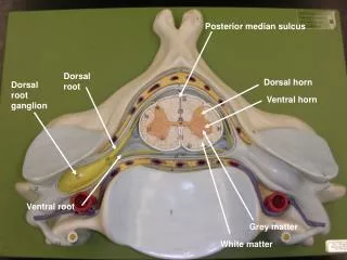
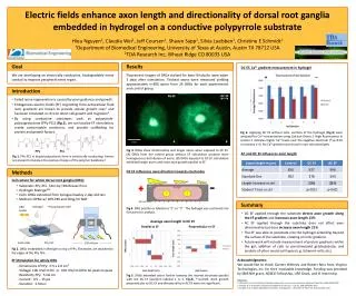
Goal • Results DC EF, Ca2+ gradient measurement in hydrogel _ _ + + • We are developing an electrically conductive, biodegradable nerve conduit to improve peripheral nerve repair. Fluorescent images of DRGs stained for beta-III-tubulin were taken 3 days after stimulation. Thickest axons were measured yielding approximately n=300 axons from 24 DRGs for each experimental and control group. * • Introduction Electric fields enhance axon length and directionality of dorsal root ganglia embedded in hydrogel on a conductive polypyrrolesubstrateHieuNguyen1, Claudia Wei1, Jeff Coursen1, Shawn Sapp2, Silvia Luebben2, Christine E Schmidt11Department of Biomedical Engineering, University of Texas at Austin, Austin TX 78712 USA2TDA Research Inc, Wheat Ridge CO 80033 USA No EF DC EF • Failed nerve regeneration is caused by poor guidance and growth. • Endogenous electric fields (EF) originating from extracellular fluid ionic gradients are known to provide cellular growth cues1 and has been simulated in vitro to direct cell growth and migration2. • By using conductive substrates such as polypyrrole-polycaprolactone (PPy-PCL) (Fig.1), we can localize EF stimulation, create customizable constructs, and provide scaffolding for protein and growth factors. Fig 6. Applying DC EF without cells, sections of the hydrogel (Fig.4) were analyzed for Ca2+ concentration using Calcium Green-1. High fluorescence in section 5 indicates higher Ca2+ levels near the negative electrode (*=p<0.01 vs sections 1-4). No Ca2+ gradient was found in non-stimulated gels. PPy PCL AC and DC EF influences axon length Fig 1. PPy-PCL in doped polycationic form is intrinsically conducting. Anions are present to balance the positive charges of the polymer backbone.3 • Methods DC EF influences axon direction towards electrodes • Cell culture for whole dorsal root ganglia (DRG) • Substrate: PPy-PCL films by TDA Research Inc • Hydrogel: MatrigelTM • Cells: DRGs extracted from Sprague Dawley 2-day-old rats • Medium: RPMI w/ 10% FBS and 50ng/ml NGF A B 1mm Fig 3. DRGs show directionality and longer axons when exposed to DC EF. (A) DRGs from the control group without EF stimulation produce more homogeneous distribution of axons. (B) DRGs exposed to DC EF stimulation exhibited longer axons and more axon growth parallel to EF. Gels sections | 1 | 2 | 3 | 4 | 5 | Hydrogel Polycarbonate well • Summary DRG bodies Fig 4. DRG position is labeled as “1” or “2”. The hydrogel was sectioned into five parts for analysis. • DC EF applied through the substrate directs axon growth along the EF gradient and increases axon length 13% • AC EF applied through the substrate does not affect axon directionality but does increase axon length 21% • The EF was able to penetrate into the hydrogel extending beyond the surface of the substrate, creating an ionic gradient. • Future work will include measurement of protein gradients within the gel, addition of cells to pre-stimulated gels/substrate, and analysis of other neural cell types (e.g. Schwann cells, etc). Average axon length in DC EF Parallel to EF Perpendicular to EF Glass slide PPy-PCL 100 mV/cm * Fig 2. DRGs embedded in Matrigel on top of PPy. Electrodes are attached to the edges of the PPyfilm. * Acknowledgments: We would like to thank Garrett Whitney and Robert Ross from Virginia Technologies, Inc. for their invaluable knowledge. Funding was provided by SBIR NIH grant, NDSEG Fellowship, URF Grant, and IE Internship. • EF Stimulation for whole DRG • - Dimensions of PPy: 0.5 x 2.0 cm2 • - Voltage: 100 mV/cm DC or 100 mV/cm 60Hz AC peak-to-peak • - Resistivity PPy: 5 kΩ·cm • - Current: 0.5 – 15 µA • - Duration: 2 hours Fig 5. DRGs extended axons further towards the nearest electrode parallel with the DC EF (position labeled 1 or 2, Fig.4), *=p<0.05. Axon growth perpendicular to DC EF and directionality in AC EF were not significant. References: 1. McCaigCD, et al. Controlling cell behavior electrically: current views and future potential. Physiol Rev. 2005;85:943-978. 2. Zhao M, et al. Orientation and directed migration of cultured... J Cell Sci. 1996;109:1405-1414. 3. Durgam H, et al. Novel degradable co-polymers of polypyrrole… J BiomaterSciPolym Ed. 2010;21:1265-1282.