Cell Culture
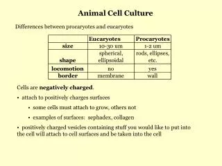
Cell Culture. Basics of Cell Culture. Cell culture is the process by which prokaryotic, eukaryotic or plant cells are grown under controlled conditions. But in practice it refers to the culturing of cells derived from animal cells.

Cell Culture
E N D
Presentation Transcript
Cell culture is the process by which prokaryotic, eukaryotic or plant cells are grown under controlled conditions. But in practice it refers to the culturing of cells derived from animal cells. Cell culture was first successfully undertaken by Ross Harrison in 1907 Roux in 1885 for the first time maintained embryonic chick cells in a cell culture Introduction
First development was the use of antibiotics which inhibits the growth of contaminants. Second was the use of trypsin to remove adherent cells to subculture further from the culture vessel Third was the use of chemically defined culture medium. Major development’s in cell culture technology
Areas where cell culture technology is currently playing a major role. Model systems for Studying basic cell biology, interactions between disease causing agents and cells, effects of drugs on cells, process and triggering of aging & nutritional studies Toxicity testing Study the effects of new drugs Cancer research Study the function of various chemicals, virus & radiation to convert normal cultured cells to cancerous cells Why is cell culture used for?
Virology Cultivation of virus for vaccine production, also used to study there infectious cycle. Genetic Engineering Production of commercial proteins, large scale production of viruses for use in vaccine production e.g. polio, rabies, chicken pox, hepatitis B & measles Gene therapy Cells having a functional gene can be replaced to cells which are having non-functional gene Contd….
Choice of media depends on the type of cell being cultured Commonly used Medium are GMEM, EMEM,DMEM etc. Media is supplemented with antibiotics viz. penicillin, streptomycin etc. Prepared media is filtered and incubated at 4 C Culture media
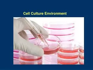
Laminar cabinet-Vertical are preferable Incubation facilities- Temperature of 25-30 C for insect & 37 C for mammalian cells, co2 2-5% & 95% air at 99% relative humidity. To prevent cell death incubators set to cut out at approx. 38.5 C Refrigerators- Liquid media kept at 4 C, enzymes (e.g. trypsin) & media components (e.g. glutamine & serum) at -20 C Microscope- An inverted microscope with 10x to 100x magnification Tissue culture ware- Culture plastic ware treated by polystyrene Basic equipments used in cell culture
If working on the bench use a Bunsen flame to heat the air surrounding the Bunsen Swab all bottle tops & necks with 70% ethanol Flame all bottle necks & pipette by passing very quickly through the hottest part of the flame Avoiding placing caps & pipettes down on the bench; practice holding bottle tops with the little finger Work either left to right or vice versa, so that all material goes to one side, once finished Clean up spills immediately & always leave the work place neat & tidy Basic aseptic conditions
Vial from liquid nitrogen is placed into 37 C water bath, agitate vial continuously until medium is thawed Centrifuge the vial for 10 mts at 1000 rpm at RT, wipe top of vial with 70% ethanol and discard the supernatant Resuspend the cell pellet in 1 ml of complete medium with 20% FBS and transfer to properly labeled culture plate containing the appropriate amount of medium Check the cultures after 24 hrs to ensure that they are attached to the plate Change medium as the colour changes, use 20% FBS until the cells are established Working with cryopreserved cells
Remove the growth medium, wash the cells by PBS and remove the PBS by aspiration Dislodge the cells by trypsin-versene Dilute the cells with growth medium Transfer the cell suspension to a 15 ml conical tube, centrifuge at 200g for 5 mts at RT and remove the growth medium by aspiration Resuspend the cells in 1-2ml of freezing medium Transfer the cells to cryovials, incubate the cryovials at -80 C overnight Next day transfer the cryovials to Liquid nitrogen Freezing cells for storage
Cells are cultured as anchorage dependent or independent Cell lines derived from normal tissues are considered as anchorage-dependent grows only on a suitable substrate e.g. tissue cells Suspension cells are anchorage-independent e.g. blood cells Transformed cell lines either grows as monolayer or as suspension Culturing of cells
Cells which are anchorage dependent Cells are washed with PBS (free of ca & mg ) solution. Add enough trypsin/EDTA to cover the monolayer Incubate the plate at 37 C for 1-2 mts Tap the vessel from the sides to dislodge the cells Add complete medium to dissociate and dislodge the cells with the help of pipette which are remained to be adherent Add complete medium depends on the subculture requirement either to 75 cm or 175 cm flask Adherent cells
Easier to passage as no need to detach them As the suspension cells reach to confluency Asceptically remove 1/3rd of medium Replaced with the same amount of pre-warmed medium Suspension cells
Human cell lines -MCF-7 breast cancer HL 60 Leukemia HEK-293 Human embryonic kidney HeLa Henrietta lacks Primate cell lines Vero African green monkey kidney epithelial cells Cos-7 African green monkey kidney cells And others such as CHO from hamster, sf9 & sf21 from insect cells Common cell lines
Cell culture contaminants of two types Chemical-difficult to detect caused by endotoxins, plasticizers, metal ions or traces of disinfectants that are invisible Biological-cause visible effects on the culture they are mycoplasma, yeast, bacteria or fungus or also from cross-contamination of cells from other cell lines Contaminant’s of cell culture
Cell viability is determined by staining the cells with trypan blue As trypan blue dye is permeable to non-viable cells or death cells whereas it is impermeable to this dye Stain the cells with trypan dye and load to haemocytometer and calculate % of viable cells - % of viable cells= Nu. of unstained cells x 100 total nu. of cells Cell viability
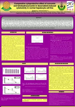
Cytotoxicity causes inhibition of cell growth Observed effect on the morphological alteration in the cell layer or cell shape Characteristics of abnormal morphology is the giant cells, multinucleated cells, a granular bumpy appearance, vacuoles in the cytoplasm or nucleus Cytotoxicity is determined by substituting materials such as medium, serum, supplements flasks etc. at atime Cell toxicity
Calcium phosphate precipitation DEAE-dextran (dimethylaminoethyl-dextran) Lipid mediated lipofection Electroporation Retroviral Infection Microinjection Transfection methods
Transfection • What is Transfection? Transfection is a method of transporting DNA, RNA and/or various macromolecules into an eukaryotic cell by using chemical, lipid or physical based methods. • Methods: (just a few examples…) MethodApplication CaPO4, DEAE DNA Transfection Liposome Based DNA Transfection Polyamine Based DNA Transfection
1928: Frederick Griffith transforms Streptococcus pneumoniae 1944: Avery, Macleod and McCarty discover DNA is the transforming factor 1964: Foldes and Trautner use the term transfection 1965: Vaheri and Pagano describe the DEAE-Dextran transfection technique Early Years • Transfection = “transformation” + “infection” • The infection of cells by the isolated nucleic acid from a virus, resulting in the production of a complete virus
Transfection vs. Transformation • Transformation: genetically altering cells by transporting in foreign genetic material. • Transfection: the process of transporting genetic material and/or macromolecules into eukaryotic cells through typically non-viral methods.
Purpose/uses of Transfection • Study gene function • Study protein expression • Transfer DNA into embryonic stem cells
How it works • Utilize Chemical, lipid or physical methods (direct microinjection, electroporation, biolistic particle delivery) for transportation of genetic materials or macromolecules.
Add chloroquine to transfection medium Add plasmid DNA to DEAE-Dextran medium Incubate cells with mixture…efficient transfection results in 25-75% death DMSO “shock” Transfection method 1: DEAE-Dextran • Facilitates DNA binding to cell membranes and entry via endocytosis • DEAE-D associates with DNAcomplex binds to cell surface
Transfection method 1: DEAE-Dextran • Major variants: number of cells, concentration of DNA and concentration of DEAE-Dextran • Only method that cannot be used for stable transfections
Transfection method 3: liposome-mediated • Use of cationic lipids • chemical and physical similarities to biological lipids • spontaneous formation of complexes with DNA, called lipoplexes • High efficiency for in vitro transfections • Can carry larger DNA than viruses • Safer than viruses • Low in vivo efficiency http://homepage.usask.ca/~sdw132/images/lipid_nanoparticle.gif
How it works – Chemical and lipid methods • Neutralize negative charges on DNA • Chemical method: Calcium phosphate; creates precipitates that settle on cells and are taken in. • Lipid or Polymer methods: interact with DNA, promotes binding to cells and uptake via endocytosis.
Experimental method/process(chemical methods) HBS = HEPES-Buffered Saline
Transfection method 2: electroporation • Use of high-voltage electric shocks to introduce DNA into cells • Cell membranes: electrical capacitors unable to pass current • Voltage results in temporary breakdown and formation of pores Harvest cells and resuspend in electroporation buffer Add DNA to cell suspension…for stable transfection DNA should be linearized, for transient the DNA may be supercoiled electroporate Selection process for transfectant
Transfection method 2: electroporation • Variants: amplitude and length of electric pulse • Less affected by DNA concentration-linear correlation between amount of DNA present and amount taken up • Can be used for stable transfections
How it works – Physical methods • Electroporation: use of high voltage to deliver nucleic acids; pores are formed on cell membrane. • Direct Microinjection: Use of a fine needle. Has been used for transfer of DNA into embryonic stem cells. • Biolistic Particle delivery: Uses high-velocity for delivery of nucleic acids and penetration of cell wall.
Advantages: Provides the ability to transfer in negatively charged molecules into cells with a negatively charged membrane. Liposome-mediated transport of DNA has high efficiency. Good for long-term studies requiring incorporation of genetic material into the chromosome. Disadvantages: Chemical Reagents: not useful for long-term studies. Transfection efficiency is dependent on cell health, DNA quality, DNA quantity, confluency (40-80%) Direct Microinjection and Biolistic Particle delivery is an expensive and at times a difficult method. Advantages/Disadvantages
Exploring protein function 1) Where is it localized in the cell? Approaches: a) Make antibodies - immunofluorescence b) “Express” the protein in cells with a tag Fuse to GFP 2) What is it doing in the cell? Approaches: a) Reduce protein levels - RNA interference b) Increase protein levels “over-express” c) “Express” mutant versions Transfection!!!!
Transfection = Introduction of DNA into mammalian cells Gene is transcribed and translated into protein = “expressed”
Direct introduction of the DNA Electroporation - electric field temporarily disrupts plasma membrane Biolistics (gene gun)- fire DNA coated particles into cell Microinjection
Infection: Use recombinant viruses to deliver DNA Retroviruses Adenoviruses Virally-mediated introduction of the DNA
Carrier-mediated introduction of the DNA Positively charged carrier molecules are mixed with the DNA and added to cell culture media: Calcium Phosphate DEAE Dextran liposomes micelles Carrier-DNA complexes bind to plasma membrane and are taken up
Types of Transfection Transient: Expression assayed 24-48 hours post transfection Stable: Integration of the transfected DNA into the cell genome - selectable marker like neomycin resistance required “stably transfected” cell line
DNA “expression” vector transfected: Insert gene in here For expression in cells Polyadenylation site GFP CMV Promoter SV40 Promoter To generate stable cell line pCMV/GFP Ampicillin resistance Neomycin resistance For amplification of the plasmid in bacteria Polyadenylation site pUC Bacterial origin of replication
Three ways to make Green fluorescent protein “GFP” fusion constructs: PROTEIN X GFP GFP PROTEIN Y PROTEIN GFP Z
EXPERIMENT: Transfect unknown GFP fusion protein Protein X, Y or Z Visualize GFP protein fluorescence by fluorescence microscopy in living cells Counter-stain with known marker to compare localization patterns inliving cells = “vital stain”
Nuclei Mitochondria Secretory Pathway: Endoplasmic Reticulum Golgi Complex Endocytotic Pathway: Endosomes • Compartments/organelles examined • Protein sequences sufficient for localization • Vital stains
Nucleus Transport through nuclear pore signal = basic amino acid stretches example: P-P-K-K-K-R-K-V
Nuclear Stain: Hoechst 33258 binds DNA