
METHODS Cell Culture
E N D
Presentation Transcript
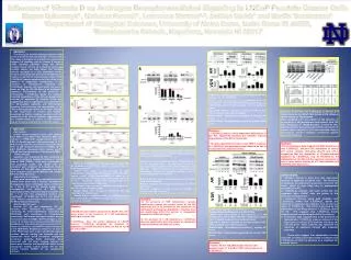
Influence of Vitamin D on Androgen Receptor-mediated Signaling in LNCaPProstate Cancer CellsMegan Rybarczyk1, Nicholas Russell1, Lawrence Mordan1,2, JoEllen Welsh1 and Martin Tenniswood11Department of Biological Sciences, University of Notre Dame, Notre Dame IN 46556, 2Kamehameha Schools, Kapalama, Honolulu HI 96817 ABSTRACT The efficacy of the anti-androgen Casodex relies on the interaction with the androgen receptor (AR). This study is designed to elucidate the interactions between AR signaling and the vitamin D receptor (VDR) signaling pathways. Using the LNCaP prostate cancer cell line as a model of early stage prostate cancer, we have shown that Casodex induces apoptosis in a time dependent manner which is co-incident with the loss of the AR from the nucleus, in agreement with previous studies. While 1,25(OH)2D3 induces apoptosis in this cell line in the absence of exogenous testosterone, in the presence of testosterone, treatment with 1,25(OH)2D3 induces cell cycle arrest but even at doses as high as 100 nM there is no evidence of apoptosis. Furthermore in the presence of testosterone, 1,25(OH)2D3 blocks the Casodex-mediated degradation of the AR and abrogates the induction of apoptosis by Casodex. These data suggest that there is significant cross talk between the AR-mediated signaling and VDR-mediated signaling that influences the susceptibility of the cells to anti-androgen induced cell death. Changes in the expression of several p73 isoforms, monitored by Western blotting, correlates with the sensitivity of the cells to Casodex, suggesting that the responsiveness of tumors to anti-androgens may be inversely related to the vitamin D3 status. AR VDR Effect of 1,25(OH)2D3 and Casodex on Nuclear AR and VDR Levels in LNCaP Cells LNCaP cells were plated in the presence of 5 nM testosterone. Cells were treated with various doses of 1,25(OH)2D3 and 100 µM Casodex for the indicated times. Top Panel:Western analysis of nuclear levels of AR, VDR, in LNCaP cells after treatment. Middle Panel: Normalized expression of nuclear AR levels Bottom Panel: Normalized expression of nuclear VDR levels. Influence of Casodex and 1,25(OH)2D3 on Nuclear p73 Splice Variants in LNCaP Cells Grown in the Absence and Presence of Testosterone Panel A:LNCaP cells were grown in the absence or presence of 5 nM testosterone and treated with 100 nM 1,25(OH)2D3, or 100µM Casodex, or both for 48h. Nuclear extracts were prepared and the expression of various splice variants of p73 was assessed using a pan-specific anti-p73 antibody normalized against lamin A/C (Panel B). METHODS Cell Culture LNCaP human prostate cancer cells obtained from ATCC (Cat # CRL-1740) (Rockville, MD) were cultured in RPMI-1640 (supplemented with 10% FBS, 1% Pen/Strept and 5 nM testosterone, at 37ºC with 5% CO2 and 95% air. LNCaP cells were treated with 25 µM etoposide dissolved in DMSO, 10-100 nM 1,25(OH)2D3 dissolved in 95% ethanol, and 10-100 µM Casodex dissolved in 75/25 ethanol:DMSO. TUNEL Analysis Apoptosis was measured by flow cytometry by terminal transferase mediated labeling of nicked ends of DNA (TUNEL). Cells were counterstained with propidium iodide, and 10,000 cells per treatment were analyzed on a Beckman Coulter Cytomics FC 500 MPL flow cytometer. The data were analyzed utilizing the Multiplus AV software (Phoenix Flow Systems). Subcellular Fractionation Cells were harvested at the indicated times. Cell pellets were lysed with a Dounce homogenizer. Homogenates were centrifuged twice at 1500 rpm for 6 min at 4ºC and the nuclear pellets were resuspended, and stored at -20ºC. The supernatants were ultracentrifuged at 55,000 rpm for 1 h at 4ºC. The cytoplasmic fraction was centrifuged at 50,000 rpm for 1 h and stored at -20ºC. Protein concentrations were determined by the Micro BCA protein assay. Western Blot Analysis Nuclear (50 g) and cytoplasmic (30 g) fractions were solubilized in loading buffer containing 5% β-mercaptoethanol, separated by SDS-PAGE, and transferred to nitrocellulose membranes. Anti-human androgen receptor mouse monoclonal (Cat # 554224; BD Pharmingen,), anti-vitamin D receptor rat monoclonal (Cat # RT-200-P0, NeoMarkers), anti-lamin A/C goat polyclonal (N-18; Santa Cruz Biotech), anti-GAPDH rabbit monoclonal (Cat # 4699-9555; Biogenesis) diluted in 5% milk in 0.05% PBS-Tween were used to identify the cognate proteins. Specific binding was detected with the appropriate horse radish peroxidase conjugated secondary antibody. Band intensities were measured with the Scion Imaging software to obtain pixel densities, and normalized relative to the appropriate loading control (Lamin A/C for the nuclear fraction; GAPDH for the cytoplasmic fraction). Summary: • 1,25(OH)2D3 exerts a dose dependent stabilization of the AR, apparently blocking the Casodex induced degradation of the AR at 72 and 96h. • The dose dependent increase in the VDR in response to 1,25(OH)2D3 has previously been shown to be due to increased transcriptional activity. Summary: •These preliminary data suggest that both testosterone and 1,25(OH)2D3influence the expression of several p73 splice variants (including ΔN-p73 and p73-ζ), suggesting that the abrogation of Casodex-induced apoptosis by 1,25(OH)2D3may be mediated by the stabilization of the AR and the selective expression of one or more splice variants of p73 that modulate p53 mediated gene expression. • CONCLUSIONS • 1. Casodex induces a dose and time dependent increase in apoptosis in LNCaP cells. The induction of apoptosis corresponds to a decrease in nuclear AR levels, which is most likely due to proteasomal degradation of AR. • 2. 1,25(OH)2D3 induces cell cycle arrest but not apoptosis when LNCaP cells are grown in the presence of testosterone. • 3. Combined therapy using both Casodex and 1,25(OH)2D3 shows that 1,25(OH)2D3 blocks the induction of apoptosis by Casodex even at very low doses of 1,25(OH)2D3. This effect is mirrored by the stabilization of the AR by doses of 1,25(OH)2D3 above 10 nM. This suggests that 1,25(OH)2D3 blunts Casodex action by stabilizing the AR in the nucleus. • Preliminary data suggests that in the presence of testosterone, 1,25(OH)2D3 induces changes in the expression of several p73 isoforms including ΔN-p73 and p73-ζ. These isoforms may modulate the induction of apoptosis through p53 mediated pathways. • These data suggest that combination therapy using the currently prescribed doses of Casodex and 1,25(OH)2D3 will NOT be effective as a treatment for prostate cancer. Effect of 1,25(OH)2D3 on the Induction of Apoptosis by Casodex in LNCaP Cells Grown in the Presence of 5 nM Testosterone. LNCaP cells, grown in the presence of 5nM testosterone, were treated with Casodex (50 or 100 µM), vehicle control (75/25 Ethanol/DMSO) or alone or in combination with 1,25(OH)2D3 for varying times from 24 to 72h. Apoptotic events were analyzed by flow cytometry as described in Methods. Top Panel: Time course of induction of apoptosis in LNCaP cells by 100 µM Casodex Middle Panel: Time course of effects of 100 nM 1,25(OH)2D3 in LNCaP cells Bottom Panel: Combined effect of 100µM Casodex and 1,25 (OH)2D3 on LNCaP cells (25µM etoposide used as positive control). AR Effect of Casodex on Androgen Receptor Levels and Subcellular Localization in LNCaP Cells Grown in the Presence of 5nM Testosterone. LNCaP cells were treated with 50 or 100 µM Casodex for the indicated times. Nuclear and cytoplasmic fractions were prepared as described in Materials and Methods. Signal intensities were quantitated and normalized. Results are reported as Mean ± SE of three independent experiments. PanelA: Nuclear levels of AR and Lamin A/C in LNCaP cells after treatment. Red line indicates the average mean density of control AR levels. Panel B: Cytoplasmic levels of AR and GAPDH in LNCaP cells after treatment. VDR Influence of Casodex on the effects of 1,25(OH)2D3 on Nuclear AR and VDR Levels in LNCaP Cells LNCaP cells were plated in the presence of 5 nM testosterone and treated 100 nM 1,25(OH)2D3 and/or 50 or 100µM Casodex for the indicated times. Top Panel:Western analysis of nuclear levels of AR, VDR, in LNCaP cells after treatment. Middle Panel : Normalized expression of nuclear AR levels. Bottom Panel : Normalized expression of nuclear VDR levels. Summary: •100 µM Casodex induces apoptosis in LNCaP cells, even when grown in the presence of 5 nM testosterone, starting as early as 24h. •1,25(OH)2D3 does not induce apoptosis in LNCaP, however 1,25(OH)2D3 abrogates the induction of apoptosis by 100 µM Casodex at doses as low as 10 nM for at least 96 h. • Summary: • In the presence of 5nM testosterone, Casodex significantly reduces the nuclear levels of the AR, apparently due to its transport to the cytoplasm and subsequent proteasomal degradation. Casodex does not significantly affect the nuclear or cytoplasmic levels of the VDR (not shown). • In the presence of 5 nM testosterone, 1,25(OH)2D3 does not significantly affect the nuclear or cytoplasmic levels of the AR or the VDR (not shown) Summary: • Neither 50 nor 100 µM Casodex influence the nuclear levels of the AR or VDR in the presence of 1,25(OH)2D3 .