Download
1 / 67
730 likes | 1.15k Vues
- Vascular permeability factors - Two main mechanisms for increased permeability of small vessels following tissue damage 1- Toxins & physical agents , cause necrosis of vascular endothelium with abnormal leakage

E N D
- Vascular permeability factors - Two main mechanisms for increased permeability of small vessels following tissue damage 1- Toxins & physical agents , cause necrosis of vascular endothelium with abnormal leakage 2- Chemical mediators , cause retraction of endothelial cells with resultant intermediate gaps
- Vascular permeability factors - Three patterns of increased leakage from vessels - Immediate transient response for 30-60 minutes mediated by histamine acting action on endothelium - Delayed response that starts 2-3 hours after injury & lasts for up to 8 hours mediated by bradykinin , factors derived from complement & factors released from dead neutrophils in exudate - Immediate response prolonged for over 24 hours if there has been direct necrosis of endothelium ( e.g. burn , chemical toxin)
- Vasoactive changes 1 - Begin with brief period of vasoconstriction - Most likely under reflex of the autonomic system sympathetic fibers around small vessels 2 - Followed by dilation of arterioles , capillaries , & postcapillary venules …….marked increase in blood flow to the affected area , sluggish flow , aids in extravasation of cells - Clinical appearance – redness & increased warmth - Vasoconstriction causes a blanching at the scratch line & then quickly disappear - Followed by vasodilation & a reddened line 3 - Erythrocytes aggregates or line up ( rouleaux)
- Post-capillary venules are primarily affected after the tissue is damaged by either : - Non-mediated mechanism - Toxins & physical agents can cause necrosis of endothelial cells resulting in abnormal leakage - Mediated ( chemical ) mechanism - Many mediators of various origins cause vasodilation & increased permeability ; Ex. Histamine - Most are short – lived
- Increased capillary permeability - Results in leakage of proteinaceous fluid ……..edema - Caused by wide spectrum of endothelial changes : - Contraction of endothelial cells in postcapillary venules with widening of interendothelial gaps - Major endothelial damage involving arterioles , capillary , & venules
- Patterns of permeability response - Biphasic response - Immediate - Very short acting ; caused mainly histamine - Delayed - Longer acting - Develops & peaks 2-4 hours after injury & lasts up to 8 hours - Mediators - Local cells – dead neutrophils - Plasma – Bradykinin & complement - Cause a widened gap junction & endothelial cell contraction
- Other possibilities - immediate – sustained or prolonged - Occurs with severe damage or injury as in a burn or toxin - Can last up to 24 hours - Delayed – sustained or prolonged - Seen in sunburns - Widened endothelial gaps
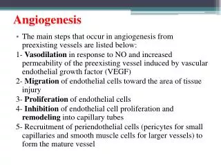
Various Mechanism Underlying Increased Vascular Permeability in inflammation - Gaps due to endothelial contraction - Direct injury - Leukocyte-dependent injury - Increased transcytosis - New blood vessel formation
Mechanism of Inflammation 1.Vasodilatation 2.Exudation -Edema 3.Emigration of cells 4.Chemotaxis
The major local manifestations of acute inflammation, compared to normal. (1)Vascular dilation and increased blood flow (causing erythema and warmth) (2)Extravasation and deposition of plasma fluid and proteins (edema) (3)leukocyte emigration and accumulation in the site of injury.
- Cellular response of leukocytes - Emigration - Adhesion molecules - Chemotaxis - Phagocytosis - Opsonization
- Cellular response of leukocytes - Emigration - Passage of inflammatory leukocytes between the endothelial cells into the adjacent interstitial tissue. - Before emigration , circulating leukocytes from the central blood move toward the endothelial surface - Margination : Leukocytes localize at outer margin of the blood flow adjacent to the vascular endothelium - Pavementing : Leukocytes line the endothelial surface - Rolling ( tumbling ) : Mediated by the action of endothelial E -& P selectins loosely binding to leukocytes & producing a characteristic “rolling” movement of the leukocytes along the endothelial surface
- Adhesion molecules - Play important role in acute inflammation - Divided into three families 1- Selectins 2- Immunoglobulin – family adhesion proteins - 3- Integrins
1- Selectins - Induced by cytokines interleukien – ( Il-1) & tumour necrosis factory ( TNF) acting on the endothelial cells - E- & P- selectins - Expressed on endothelial cells & bind to oligosaccharides ( e.g. Slalyl – lewis X) on the surface of leukocytes - P-selectins are stored in Weibel - Palade bodies & platelet alpha granules ….. Relocate to the plasma membrane after stimulation by histamine & thrombin - L-selectins - Expressed on neutrophils & bind to endothelial mucin-like molecules ( e.g. Glycam-1)
2- Immunoglobulin – family adhesion proteins - Intercellular adhesion molecules 1& 2 ( ICAM-1 & ICAM-2 ) expressed on endothelial cells & bind to integrin molecules on leukocytes - Vascular adhesion molecules ( VCAMs) similarly are expressed on endothelial cells & bind to integrin molecules on leukocytes 3- Integrins - Ex. – leukocyte LFA-1 , MAC-1 & VLA -4 - Bind to immunoglobulin – family adhesion proteins
- Emigration – Cont - Adhesion / aggregation - Leukocytes adhere to endothelial surface & aggregate to each other - Mediated by interaction of integrins on leukocytes binding to immunoglobulin-family adhesion proteins on endothelium ( activated) - IL-1 & TNF activate endothelial receptors of the smallest vessels - Transmigration - Movement of leukocytes across the endothelium - Mediated by platelet endothelial cell adhesion molecule 1 (PECAM -1) on both leukocytes & endothelium
- Three general mechanisms Mediating increased leukocyte – endothelial adhesion in the setting of inflammatory stimuli A- Redistribution of P- selectin B- Cytokine activation of endothelium , resulting in increased de novo synthesis of adhesion molecules C- Incraesed binding affinity of integrins with ICAM-1 , intercellular adhesion molecule 1; IL-1, interleukin 1 , TNF , tumour necrosis factor
Sequence of Events in Inflammatory leukocyte Emigration The leukocytes : 1- Roll 2- Adhere to endothelium 3- Transmigrate through an intercellular junction & pierce the basement membrane 4- Migrate toward chemoattractants released from a source of injury
- Chemotaxis - Process by which leukocytes are attracted to & move toward injury ( unidirectional locomotion) - Neutrophils , macrophages & lymphocytes - Mediated by diffusible chemical agents ( factors) - Movement of leukocytes occurs along a chemical gradient of factors - Receptors detect the chemical concentration gradient - Measured in an in vitro system – Boyden chamber - Assesses migration of cells from an upper chamber through a microporous membrane to a lower chamber filled with a chemoattractants
- Chemotaxis - Chemotactic factors for neutrophils produced at the site of injury - Products from bacteria - Complement components , especially C5a - Arachidonic acid metabolites , especially : - Leukotriene B4 ( LTB4) - Hydroxyeicosatetraenoic acid ( HETE) - Kallikrein
Chemotactic Factors or mediators - Products of bacteria , viruses , cells - Activated or released products of inflammation - Complement components – C3, C5,C6,C7 - Fibrin & plasmin systems - Collagen - Leukotrienes
- Cellular response of leukocytes - Phagocytosis - Ingestion & processing of particulate material by phagocytic cells. - Tissue debris , living or dead bacteria , other foreign cells - Neutrophils & monocytes-macrophages are the most important phagocytic cells.
- Cellular response of leukocytes - Phagocytosis - Process - Adherence /Binding - Matter to cell surface - Aided by opsonization - Ingestion - Matter to form phagocytic vacuole or phagosome - Fusion - Phagosome with lysosome to form phagolysosome ( PH=4) - Killing occurs inside the phagolysosome - Degradation -results in residual body or extrusion
Phagocytosis of a particle - attachment ( adherence) & binding of opsonins ( e.g. , collectins , or C3b & Fc portion of immunoglobulin ) to receptors on the leukocyte surface - Engulfment ( ingestion) - Fusion of the phagocytic vacuole with granules ( lysosomes) , & degranulation ( degradation)
- Cellular response of leukocytes - Opsonization - “ to prepare the table - Coating of particulate material - Immunoglobulin ( Ig) or - Complement ( C) - Facilities phagocytosis – “ adherence /binding” step
- Opsonization - IgG & C3b cellular receptors on membranes of neutrophils & macrophages bind firmly to IgG & C3b molecules which are attached to particles or cells - Binding immobilizes the particles on the surface of the phagocyte ( i.e. neutrophils & macrophages) - Fragment opsonized by IgG is bound to phagocytic cells by cell surface receptors for the Fc portion of the IgG molecule - Fragments opsonized by C3b are bound to cellular receptors for C3b
Cellular response of leukocytes - Phagocytosis - Anatomic changes - Internalization of attached opsonized particle by pseudopodial extensions from the surface of the leukocyte - Enclose the foreign particle ………phagosome ( internalized vesicle) - Phagosomes fuse with cytoplasmic lysosomes …… phagolysomes - Phagolysome formation associated with leukocytic degranulation
The lymphatics in Acute Inflammation - Lymphatic channels become dilated & drain the edema fluid or exudate with it is large proteins from the inflammatory site - Drainage limits the swelling - If blocked , severe edema results = lymphedema - Early in the inflammatory process - Lymphatics by fibrin - Later in the inflammatory process - Plasmin system breaks down the fibrin & opens up the lymphatic drainage
Hereditary Defects that impair the Acute Inflammatory Response - Deficiency of complement components - Increased susceptibility to infection - Particularly complement components - C2 - C3 - C5
- Defects in neutrophils - Chronic granulomatous disease of childhood - Usually X-linked with deficient activity of NADPH oxidase - Phagocytic cells ingest but do not kill certain microorganisms - Catalase-positive organisms - Ingested but not killed e.g. Staph aureus , can destroy H2O2 generated by bacterial metabolism & so H2O2 not available for myeloperoxidase – halide - Catalase – negative organisms - Ingested & killed e.g , strep , that produce sufficient H2O2 to permit oxygen –dependent microbicide so the bacteria in a sense kill themselves
Defects in neutrophils - Myeloperoxidase deficiency - Sometimes recurrent infections but often no clinical consequences - Chediak – Higashi syndrome - Autosomal recessive - Neutropenia , albinism , cranial & peripheral neuropathy , repeated infections - Abnormal WBC : - Functionally have abnormal microtubule formation affecting movement so impaired chemotaxis & migration - Morphologically have large cytoplasmic granules ( abnormal lysosomes) in granulocytes , lymphocytes , & monocytes & large melanosomes in melanocytes
Leukocyte Function Defects - Causes of deficient phagocytosis - Opsonization defects - Hypogammaglobulinemia - Engulfment defects - Drugs ; morphin - Degranulation defects - Drugs , steroids - Lack of NADPH-oxidase & failure to kill organisms - Severe glucose -6-phosphate dehydrogenase deficiency - No peroxide is generated
Outcome of the acute inflammatory reaction - Depends upon the removal of the inflammatory exudate & it is replacement either by regenerated cells of the original type ( restitution ) or by scar tissue ( fibrous repair ) - End result : - Resolution - Healing by repair - Chronic inflammation
- Resolution 1 - The best possible outcome not common & usually supporting stroma damage & healing is by scar formation 2 - Occurs when even through extensive destruction damage to the supporting tissues is minimal 3 - Acute inflammation exudate resolves & only the epithelial cells are deficient a - Exudate removed by liquefaction by neutrophil enzymes & phagocytosis of particulate debris by macrophages b - Damaged cells regenerate leaving the structure virtually as before & normal function regained c- Ex ample – lobar pneumonia
Factors favoring resolution : 1- Minimal cell death 2- Tissue has regenerative capacity 3- Fast destruction of agent 4- Fast removal of debris 5- Good drainage
-Resolution key facts : 1- End result is restoration of normal structure & function without scarring 2- Acute inflammatory exudates removed by liquefaction & phagocytosis 3- Support stroma must be intact 4- Damaged cells must be able to regenerate - Only certain cells can regenerate a- Surface epithelia constantly dividing b- Liver & kidney can divide if to replace after damage c- Nerve & cardiac cells cannot divide so loss is permanent
I ) Labile cells a- High turnover rate b- Have high capacity for regeneration c- Ex. – squamous , glandular , & GI epithelium ; bone M. II) Stable cells a- Low turnover rate b- Can proliferate rapidly when called upon c- Ex. Liver , kidney , fibroblasts , osteoblasts & endothelial III) Permanent cells a- cannot divide b- Cannot regenerate c- Heal with scar tissue d- Ex. Neurons & cardiac cells
- Organization & repair 1- Type of healing that occurs when substantial structural damage to the tissue stroma 2- Eventually leads to formation of a scar 3- Sequence of changes : - Pre-existing capillaries in undamaged tissue form new capillaries by budding into the damaged area which is also infiltrated by macrophages , fibroblasts , & myofibroblasts a- Macrophages phagocytize inflammatory exudate & dead tissue b- Vascular granulation tissue ( fragile complex of interconnecting capillaries , macrophages , & support cells) replaces the area of tissue damage.
-Organization & repair - Sequence of changes : 1- Progressive growth of fibroblasts & myofibroblasts & tissue defect filled with complex capillary network & a few residual macrophages ( fibrovascular granulation tissue) - Many of the newly formed capillaries regress until only a relative few vascular channels remain 2- Intervening spaces between vessels come progressively filled with fibrous granulation tissue a- Fibroblasts align themselves so collagen deposited in a uniform pattern that follows a direction best adapted to physical stresses b- Contraction of the granulation occurs ( myofibroblasts) & thus the size of the damaged area is reduced 3- Production of dense collagen by fibroblasts forms a collagenous scar