Download
1 / 1
10 likes | 146 Vues
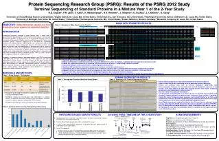
Settled on 3 standard proteins for distribution as separated proteins in Year 1 of the study. ABRF 2012. ABRF 2011. Extended deadline for returning data. Aug ‘ 11. Mar ‘12. Feb ‘13. Jan ‘ 12. Feb ‘ 11. Sep ‘ 11. Data analysis. May ‘ 12. Oct ‘11. Samples sent

E N D
Settled on 3 standard proteins for distribution as separated proteins in Year 1 of the study ABRF 2012 ABRF 2011 Extended deadline for returning data Aug ‘11 Mar ‘12 Feb ‘13 Jan ‘12 Feb ‘11 Sep ‘11 Data analysis May ‘12 Oct ‘11 Samples sent to participants Year 1 (2012) Study announcement May ‘11 Discussed ideas for 2012 study. Agreement upon a study design. Protein Sequencing Research Group (PSRG): Results of the PSRG 2012 Study Terminal Sequencing of Standard Proteins in a Mixture Year 1 of the 2-Year Study R.D. English1, P.R. Jalili2, V. Katta3, K. Mawuenyega4 , H.A. Remmer5, J. Simpson6, D. Suckau7, J.J. Walters2 , B. Xiang8 1University of Texas Medical Branch, United States, 2Sigma-Aldrich, St. Louis, MO, United States,3Genentech Inc., San Francisco, CA, United States, 4Washington University School of Medicine, St. Louis, MO, United States, 5University of Michigan, Ann Arbor, MI, United States, 6United States Pharmacopeia, Rockville, MD, United States, 7Bruker Daltonics, Bremen, Germany, 8Monsanto Company, St. Louis, MO, United States MASS SPECTROMETRY RESULTS OBJECTIVE:Obtain N-terminal sequence of three standard proteins supplied as separated samples. INTRODUCTION Establishing N-terminal sequence of intact proteins plays a critical role in biochemistry and drug development. N-terminal sequence analysis is necessary for QC of protein biologics, for determining sites of signal peptide cleavage events, for characterizing monoclonal antibodies, and in elucidating sequences of genes from uncommon species. N-terminal sequencing is in the midst of a technology transition from classical Edman sequencing to mass spectrometry (MS) based terminal sequencing. Protein homogeneity (absence of interfering protein, non-protein contaminants and buffer components) is absolutely critical to the success of analysis. While it has been very straightforward to isolate the protein of interest out of protein mixtures in preparation for Edman sequencing, core facilities now need to adopt novel sample preparation techniques to isolate proteins in high purity and make them amenable for terminal sequencing by MS. There is a lack in easy-to-use, field proven methods for sample clean-up. To address the upcoming change in technology platforms, the PSRG is conducting atwo-yearstudy with the ultimate goal of sample preparation and terminal sequencing of a protein mixture. This year’s study is the first phase towards the overall goal and entails terminal sequencing and identification of (fusion) proteins, which were provided as separated, homogeneous proteins. Participants used Edman and/or MS techniques, along with bioinformatics tools to derive the termini of the sample proteins. Study participants were directed to a website to anonymously upload sequences and data. MATERIALS AND METHODS Table 3. Summary of Mass Spectrometry Results • Top-Down with ETD or ISD provides reliable N-term sequences. • Edman and Top-Down complement each other very well: Edman for the first ~10 residues, Top-Down for the inexpensive extension of calls. • Edman was replaced successfully in validating the N-term sequence by either T³-sequencing or Bottom-Up work. • Successful analysis of the fusion Protein A required greater user expertise. • Top-Down CID provided the least robust assignments and were most easily misinterpreted. • Top-Down by ETD or ISD enabled the detection of the C-terminal removal of lysine and intact MW determination validated these findings. • Ragged N-termini in case of Endostatin were recognized by those that determined the intact molecular weight(s) or that used Edman. • Use of protein HPLC allowed the separation of Endostatin fragments but resulted in shortened readouts. • Bottom-Up alone did not assign the termini correctly, and uncertainty in assigning sequences near Protein A fusion site caused incorrect assignments. Table 1. Study Design – The Samples EDMAN DEGRADATION RESULTS Table 4. Average # of Residues Identified Using Edman Table 5. Summary of N-Terminal Sequencing Results • Edman sequencing allows for direct determination of the N-term sequence. • Labs returned N-term data correlating well with published protein sequences. • Edman can produce data with or without separation (SDS-PAGE and chromatography). However, the lower abundant second Endostatin sequence in the mixture was more difficult to deal with, causing shorter read length, erroneous assignments, and 2 participants failed to detect it. • No C-term data were produced with Edman. • The N-terminus (methylated Met-1) in protein A was not detected, but enzymatic and chemical methods allowed to obtain correct sequences from residue 2 onwards. Calling the correct ß-glucuronidase sequence was straightforward for all that used Edman while a correct Top-Down sequencing result depended much more on the right “work-hypothesis” to be successful and additional validation efforts. • Some Edman participants reported an N-term Phe (F) or an N-term unknown amino acid (X), rather than the met-M, presumably because of possible co-elution of PTH Phe and PTH Methyl-Met on Edman sequencers (under investigation while poster in print). • All Participants used ABI instrumentation, with the exception of Participant A00 who used a Shimadzu product. • Participants were asked to analyze the samples for terminal sequencing using any technology available. • Participants received all three proteins with identification in sufficient amounts to sequence each protein utilizing all three technologies. Feasibility of analysis had been previously validated by PSRG members. • Participants also filled out a survey, all responses were kept anonymous. Table 2. Instrumentation used by Participating Laboratories PARTICIPATION AND SURVEY RESULTS 2012/2013 PSRG: TIMELINE OF THE 2-YEAR STUDY ACKNOWLEDGEMENTS RepliGen for the generous gift of rec. Protein A. Sigma for the generous gift of Endostatin. Xuemei Luo (University of Texas Medical Branch) for data accumulation and anonymity. Steve Smith (University of Texas Medical Branch) and Larry Dangott (Texas A&M University) for Edman sequencing to provide reference data for this study. Anja Resemann (Bruker Daltonics) for Top-Down MS sequencing to provide reference data for this study. The ABRF Executive Board for support and scrutiny of study proposal. All Participating Labs for analyzing samples and returning data. • 25 laboratories from 12 countries requested samples for Edman sequencing and most of the labs (23) also for MS sequencing. • 14 of the 25 participating laboratories (56%) completed the survey. • 7 of the 14 labs utilized Edman sequencing , 6 top-down MS and 1 bottom-up MS (5 used bottom-up for confirmation). • Out of 14 respondents, • 9 labs analyzed the reference protein BSA, 8 correctly determined the N-terminus. • 13 labs analyzed Protein A , 4 correctly determined the N-terminus (methyl-Met). • 14 labs analyzed Endostatin, 12 labs correctly determined the N-terminus , only 7 identified the presence of the second N-terminus. Deadline for returning data ABRF 2013 Oct ‘12 Data analysis Jun ‘12 Distribution of proteins in mixture for year 2 of the study Year 2 (2013) Study announcement