Understanding the Mediastinum: Structures and Functions
Explore the divisions and contents of the mediastinum, including details on pleura, lungs, and applied anatomy. Learn about the blood supply and nerve distribution in this anatomical region.

Understanding the Mediastinum: Structures and Functions
E N D
Presentation Transcript
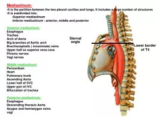
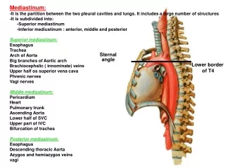
Mediastinum: -It is the partition between the two pleural cavities and lungs. It includes a large number of structures -It is subdivided into: -Superior mediastinum -Inferior mediustinum : anterior, middle and posterior Superior mediastinum: Esophagus Trachea Arch of Aorta Big branches of Aortic arch Brachiocephalic ( innominate) veins Upper half os superior vena cava Phrenic nerves Vagi nerves Middle mediastinum: Pericardium Heart Pulmonary trunk Ascending Aorta Lower half of SVC Upper part of IVC Bifurcation of trachea Posterior mediastinum: Esophagus Descending thoracic Aorta Azygos and hemiazygos veins vagi ِ Sternal angle angle Lower border of T4
Pleura: • -Completely close sac invaginated by the • lung from the medial side • -Parietal pleura and visceral pleura • Subdivision of parietal pleura • 1- cervical • 2- Costovertebral • 3- mediastinal • 4- diaphragmatic
Lines of pleural reflections: 0, 2, 4, 6, 8, 10 and 12 Blood supply= thoracic wall Nerve supply= lungs
Lungs: Apex, base, costal surface, medial surface 3 borders; anterior, inferior and posterior
Mediastinal surface Hilum containing the root of the lung Vertebral surface Hilum of the lung:It is the part of the mediastinal surface which gives passage to the structures forming the root of the lung Root of the lung:1- Bronchus (Rt, Lt) 2- Pulmonary artery (superior) 3- Pulmonary veins (sup., inf.) 4- Pulmonary nerve plexus 4- Lymph nodes 5- Bronchial vessels (Aorta, Azygos)
Lobes and fissures of lungs: Left lung:Oblique fissure 21/2 inches below apex opposite 3rd Th.V. till inferior border opposite 6th Costochondral junction 2 lobes: superior and inferior Lobes and fissures of lungs: Right lung:Oblique fissure Horizontal fissure: from ant. border opposite 4th costal cartilage till oblique 3 lobes: sup., middle and inf. Differences between Rt, Lt lungs: 1- size and weight 2- length and breadth 3- anterior border 4- hilum How to identify the right and left lungs?
Surface anatomy of the lungs: 0, 2, 4, 6, 6, 8, 10 Applied Anatomy: -Bare area of pericardium -Pericardial puncture -Stab wound in the midaxillary line .below 10th rib . between 8-10 . above 8th rib -Pneumothorax -Hydrothorax