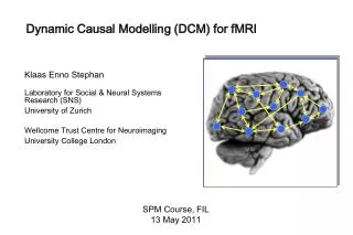
DCM: Dynamic Causal Modelling for fMRI
Wellcome Trust Centre for Neuroimaging. SPM-Course October 2013. DCM: Dynamic Causal Modelling for fMRI. Mohamed Seghier Wellcome Trust Centre for Neuroimaging , University College London, UK. ?. ?. ?. Functional integration : How do regions influence each other?

DCM: Dynamic Causal Modelling for fMRI
E N D
Presentation Transcript
Wellcome Trust Centre for Neuroimaging SPM-Course October 2013 DCM: Dynamic Causal Modelling for fMRI Mohamed Seghier Wellcome Trust Centre for Neuroimaging, University College London, UK
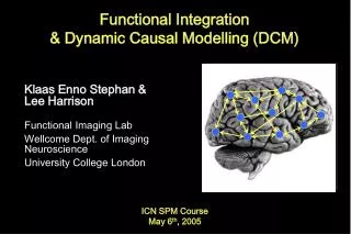
? ? ? Functional integration: How do regions influence each other? Brain Connectivity Functional segregation: What regions respond to a particular experimental input?
Connectivity is an important facet of brain function: • ** Regions don’t operate in isolation ** Neurodegenerative and psychiatric disorders = a disorder of brain connectivity. E.g.: Schizophrenia and autism [Smith 2012 Nature]
anatomical/structural connectivity= presence of axonal connections. • functional connectivity= statistical dependencies between regional time series. • effective connectivity= causal (directed) influences between neurons or neuronal populations. [Sporns 2007, Scholarpedia]
Structural connectivity - Presence of axonal connections: relay and coordinate communication between different brain regions The function of the axon is to transmit information to different neurons c.f. Els Fieremans Dissected white matter - E.g. measured with tracing techniques or diffusion tensor/spectrum imaging (DTI/DSI)
Knowing anatomical connectivity is not enough... • Connections are recruited in a context-dependent fashion: • Local functions depend on network activity • Connections show synapticplasticity • Critical for learning • Can occur both rapidly and slowly Need to look at functional/effective connectivity. But: ** Anatomo-functional connectivity: combine functional with structural connectivity.
Functional connectivity = statistical dependencies (temporal correlations) between activations. seed region [Biswal et al. 1995 MRM] - Seed-based correlation analysis - Coherence analysis - Eigen-decomposition (e.g. SVD) - Clustering (e.g. FCM) - Independent component analysis (ICA)
♣Whole-brain regression with seed regions: functional connectivity maps seed region ♦ Controlled task: reading words, pseudowords, letter strings. [Bokde et al. 2001 Neuron] ♦ Uncontrolled task (= unlocked onsets): continuous sentence reading. [Hampson et al. 2006 Neuroimage] Seed ROI = left angular gyrus. Functional connectivity maps vary during (natural) reading of sentences. Seed ROI = left inferior frontal gyrus. Functional connectivity maps vary with word type. E.g. watching movies / sleep / hallucinations
Pros & Cons of functional connectivity analysis • ** Pros: • - Easy to compute; • - useful when we have no experimental control over the system of interest and no model of what caused the data (e.g. sleep, hallucinations, natural vision). • ** Cons: • - interpretation of resulting patterns is difficult / arbitrary; • - no mechanistic insight. Effective connectivity
fMRI experiment; task contrasts Effective connectivity Can we go beyond this “static” picture? Dynamics or interactions between regions… For understanding brain function mechanistically, we need models of effective connectivity, = causal (directed) influences between neurons or neuronal populations. explain regional effects in terms of interregional connectivity.
FMRI response = indirect + slow [Arthurs & Boniface 2002 TINS] parameterise effective connectivity in terms of coupling among unobserved brain states (e.g., neuronal activity in different regions). Neuronal: Unobserved interactions BOLD: Measured responses DCM ** simple neuronal model; ** complicated hemodynamic forward model (neural activity BOLD).
[Friston et al. 2003 Neuroimage] The hemodynamics Deterministic dynamical systems [Friston 2002 Neuroimage] [Friston et al. 2000 Neuroimage]
DCM is a generative model = a quantitative/mechanistic description of how observed data are generated/caused. Key features: 1- Dynamic 2- Causal 3- Neuro-physiologically motivated 4- Operate at hidden neuronal interactions 5- Bayesian in all aspects 6- Hypothesis-driven 7- Inference at multiple levels. [Stephan et al. 2010 Neuroimage] DCM [default] implementation: Deterministic Stochastic[Daunizeau et al. 2009] Bilinear Nonlinear[Stephan et al. 2008] The one-state neuronal The two-state[Marreiros et al. 2008]
z λ y Basic idea of DCM for fMRI ♣ A cognitive system is modelled at the neuronal level (not directly accessible for fMRI). ♣ The modelled neuronal dynamics (z) is transformed into area-specific BOLD signals (y) by a hemodynamic forward model (λ). Aim:to estimate the parameters of a reasonably realistic neural model such that the predicted/modelled BOLD responses correspond as closely as possible to the observed/measured BOLD responses.
What is a system? System = a set of elements which interact in a spatially and temporally specific fashion Input u(t) connectivity parameters system states z(t) • State changes of a system are dependent on: • the current state z • external inputs u • its connectivity q (evolution equation)
u1 u2 R1 z1 z2 R2 activity in is coupled to via coefficient Neurodynamics: 2 nodes with input
u1 u2 R1 z1 z2 R2 modulatory input u2 activity through the coupling Neurodynamics: positive modulation
u1 u2 z1 z2 reciprocal connection disclosed by u2 Neurodynamics: reciprocal connections
u1 u2 CONTEXT u3 R1 left R4 right R2 right R3 left bilinear dynamic system z4 z3 z1 z2
Bilinear state equation in DCM for fMRI The neural state equation modulation of connectivity state vector direct inputs state changes externalinputs connectivity n regions m inputs (driv.) m inputs (mod.)
“C”, the direct or driving effects: - extrinsic influences of inputs on neuronal activity. • “A”, the endogenous coupling or the latent connectivity: • - fixed or intrinsic effective connectivity; • first order connectivity among the regions in the absence of input; • average/baseline connectivity in the system. • “B”, the bilinear term, modulatory effects, or the induced connectivity: • context-dependent change in connectivity; • - eq. a second-order interaction between the input and activity in a source region when causing a response in a target region. [Units]: rates, [Hz]; Strong connection = an effect that is influenced quickly or with a small time constant.
B A DCM parameters = rate constants Integration of a first-order linear differential equation gives an exponential function: z1 Decay function 1 0.8 If AB is 0.10 s-1 this means that, per unit time, the increase in activity in B corresponds to 10% of the activity in A 0.6 0.4 0.2 0.10 0 -0.1 0 0.1 0.2 0.3 0.4 0.5 0.6 0.7 0.8 0.9
modulatory input u2(t) driving input u1(t) t t y BOLD y y y hemodynamic model λ activity x2(t) activity x3(t) activity x1(t) z neuronal states integration Neuronal state equation The bilinear model endogenous connectivity modulation of connectivity [Stephan & Friston (2007),Handbook of Brain Connectivity] direct inputs
t u inputs The hemodynamic model neural state equation • Hemodynamic parameters: neuronal input z(t) important for model fitting, but of no interest for statistical inference hemodynamic state equations • Empirically determineda priori distributions. • Area-specific estimates (like neural parameters) region-specificHRFs !! Balloon model BOLD signal y(t) BOLD signal change equation [Friston et al. 2000, NeuroImage] [Stephan et al. 2007, NeuroImage]
u2 u1 R1 left R2 right R4 right R3 left Example: modelled BOLD signal Recap: The aim of DCM is to estimate: • Neuronal parameters [A, B, C]; • Hemodynamic parameters; • Such that modelled/predicted and measured/observed BOLD signals are maximally similar. Multiple-input multiple-output system black: observed BOLD signal red: modelled BOLD signal
The posterior probability of the parameters given the data is an optimal combination of prior knowledge and new data, weighted by their relative precision. Priors in DCM Constraints on parameter estimation: • - hemodynamic parameters: empirical priors • - coupling parameters other connections: shrinkage priors Priors & parameter estimation Based on a Bayesian framework. Bayes theorem allows us to express our prior knowledge or “belief” about parameters of the model. new data prior knowledge posterior likelihood ∙ prior
Inference about DCM parameters: Bayesian inversion • Gaussian assumptions about the posterior distributions of the parameters (mean ηθ|y and covariance Cθ|y). • Use of the cumulative normal distribution to test the probability that a certain parameter (or contrast of parameters cTηθ|y) is above a chosen threshold γ: • By default, γ is chosen as zero ("does the effect exist?"). ηθ|y ** Parameter estimation by means of VariationalBayesunder the Laplace approximation scheme (VL). [Friston et al. 2007 Neuroimage]
Modulatory input (e.g. context/learning/drugs) Driving input (e.g. sensory stim) b12 c1 c2 a12 ηθ|y y BOLD y DCM: practical steps Select areas you want to model • Extract timeseries of these areas (x(t)) • Specify at neuronal level • what drives areas (c) • how areas interact (a) • what modulates interactions (b) • State-space model with 2 levels: • Hidden neural dynamics • Predicted BOLD response • Estimate model parameters: Gaussian a posteriori parameter distributions, characterised by mean ηθ|y and covariance Cθ|y. neuronal states activity x1(t) activity x2(t)
Attention to motion in the visual system Stimuli 250 radially moving dots at 4.7 degrees/s Pre-Scanning 5 x 30s trials with 5 speed changes (reducing to 1%) Task - detect change in radial velocity Scanning(no speed changes) 6 normal subjects, 4 x 100 scan sessions; each session comprising 10 scans of 4 different conditions F A F N F A F N S ................. F - fixation point only A - motion stimuli with attention (detect changes) N - motion stimuli without attention S - no motion Attention – No attention [Büchel & Friston 1997, Cereb. Cortex] [Büchel et al.1998, Brain]
Choice of areas and time series extraction. Three ROIs: V1, V5, and SPC. Definition of driving inputs. All visual stimuli/conditions (photic: A N S) Definition of modulatory inputs. The effects of motion and attention (A N) Building the model: 1- how to connect regions (intrinsic connections “A”); 2- how the driving inputs enter the system (extrinsic effects “C”); 3- define the context-dependent connections (modulatory effects “B”). How we can interpret, mechanistically, the increase in activity of area V5 by attention when motion is physically unchanged. SPC V5 Attention – No attention
SPC V1 V5 • Visual inputs drive V1. • Activity then spreads to hierarchically arranged visual areas. • Motion modulates the strength of the V1→V5 forward connection. • Attention modualtes the strength of the SPC→V5 backward connection. Attention Motion Photic Re-analysis of data from[Friston et al., 2003 NeuroImage]
Attention Re-analysis of data fromFriston et al., NeuroImage 2003 SPC V1 V5 0.37 0.56 0.42 Motion 0.66 0.88 -0.05 Photic 0.48 After DCM estimation: • Motion modulates the strength of the V1→V5 forward connection. • Attention increases the backward-connection SPC→V5. Are there other plausible/alternative models?
Model 1:attentional modulationof V1→V5 Model 2:attentional modulationof SPC→V5 Model 3:attentional modulationof V1→V5 and SPC→V5 SPC SPC V1 V1 V5 V5 Attention Attention Photic Photic Photic SPC 0.55 0.03 0.85 0.86 0.85 0.70 0.75 0.70 0.84 1.36 1.42 1.36 0.89 0.85 V1 -0.02 -0.02 -0.02 0.56 0.57 0.57 V5 Motion Motion Motion 0.23 0.23 Attention Attention Alternative models (hypothesis-driven approach): How we can compare between competing hypotheses? BMS (Bayesian Model Selection)
[Pitt and Miyung 2002 TICS] Model evidence and selection Given competing hypotheses, which model is the best? Which model represents thebest balance between model fit and model complexity? For which model m does p(y|m) become maximal?
The negative variotional free energy (F) approximation Under Gaussian assumptions about the posterior (Laplace approximation), the negative free energy Fis a lower bound on the log model evidence. Approximations to the model evidence in DCM Log model evidence = balance between fit and complexity. [Penny 2012, NeuroImage] - A better approximation of the complexity term: Faccounts for parameter interdependencies. ** All recent DCM versions use F for model selection !
Inference on model space BMS (Bayesian Model Selection) An intuitive interpretation of model comparisons is made possible by Bayes factors: Model m2 Model m1 positive value, [0;[ [Kass & Raftery 1995, J. Am. Stat. Assoc.] !!# Only compare models with the samedata #!!
BMS has nothing to say about the “true” model(s). find the most useful model, form a set of alternatives, given data. Best model = best balance between accuracy and complexity. • - model selection with BMS model validation! DCM model space: Compatibility // Size // Plausibility. # BMS cannot be applied to models fitted to different data! (Only models with the same ROIs can be compared using BMS). • # It is helpful to constrain your DCM model space.number of ROIs limited to 8 in SPM (GUI), but you can include more ROIs.(e.g., 6 ROIs, fully connected, 1 Billion alternative modulations!). • # (if possible) Define sets of models that are plausible, in a systematic way, given prior knowledge (e.g. anatomical, TMS, previous studies). • # for group comparison (e.g. patients vs. controls) make inferences over the same DCM model space.
Levels of inference: Group level -- Family level -- -- System/model level -- -- Parameter/connection level -- FFX: subjects assumed to use similar systems. RFX: best models vary across subjects. [Penny et al. 2010, PLoS Comp Biol] [Seghier et al. 2010, Front SystNeurosci] • ♣ Family level: • - Useful when no clear winning model // models have common characteristics. Models assigned to subsets (families) with shared features. • Inference: a class/type of models that best explains the data. • ♣Model level: • - Useful when a clear winning model can be identified (BMS). • Inference: a useful model structure (inputs & connections) that explains the data. • ♣ Connection level: • - Useful when connectivity parameters are of interest (e.g. modulations). • Inference: Bayesian parameters averaging (BPA) or t-test on DCM parameters. • Inference: BMA on the winning family (or over the whole model space).
Which DCM version? DCM5 || DCM8 || DCM10 || DCM12. • Use the latest version (= DCM12). • Keep the same DCM version for your project (over models, sessions, and subjects). • - Indicate the DCM version in your papers. Extensions in DCM for fMRI: • Bayesian Model Selection BMS [Penny et al. 2004 Neuroimage]. • Slice specific sampling [Kiebel et al. 2007 Neuroimage]. • Refined hemodynamic model [Stephan et al. 2007 Neuroimage]. • The two-state DCM [Marreiros et al. 2008 Neuroimage]. • The non-linear DCM [Stephan et al. 2008 Neuroimage]. • Random-effects BMS [Stephan et al. 2009 Neuroimage]. • Stochastic DCM [Daunizeau et al. 2009 Physica D]. • Anatomical-based priors for DCM [Stephan et al. 2009 Neuroimage]. • Family level inference BMS [Penny et al. 2010 PLoS Comp Biol]. • Bayesian model averaging BMA [Penny et al. 2010 PLoS Comp Biol]. • Post-hoc Bayesian optimisation [Friston et al. 2011 Neuroimage]. • Stochastic DCM (random fluctuations)[Li et al. 2011 Neuroimage]. • Network discovery for large DCMs [Seghier & Friston et al. 2013 Neuroimage].
Reviews: Stephan et al. (2010). Ten simple rules for DCM. NeuroImage. Daunizeau et al. (2010).DCM: a critical review of the biophysical and statistical foundations. NeuroImage. Seghier et al. (2010).Identifying abnormal connectivity in patients using dynamic causal modeling of fMRI responses . Front Syst Neurosci. Friston (2011). Functional and effective connectivity: A review.Brain Connectivity. Practical examples: (DCM-fMRI at the FIL) - Inter-hemispheric interactions and laterality for words and pictures: Seghier et al. (2011) Cerebral Cortex. - Prediction error and putamen: den Ouden et al. (2010) J Neurosci. - Top-down effects on form perception: Cardin et al. (2011) Cerebral Cortex. - Multilingual vs. Monolingual monitoring of speech production: Parker-Jones et al. (2013) J Neurosci. http://www.fil.ion.ucl.ac.uk/spm/data/
Wellcome Trust Centre for Neuroimaging SPM-Course October 2013 Thank you for your attention!!!