Reflex Physiology
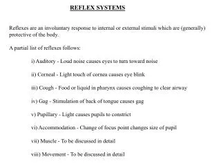
Reflex Physiology. Reflexes are automatic, unconscious to changes, either inside or outside the body. Reflexes maintain homeotasis (autonomic reflexes) – heart rate, breathing rate, bp , digestion. Reflexes also carry out the automatic actions of swallowing, sneezing, coughing, vomiting.

Reflex Physiology
E N D
Presentation Transcript
Reflexes are automatic, unconscious to changes, either inside or outside the body. • Reflexes maintain homeotasis (autonomic reflexes) – heart rate, breathing rate, bp, digestion. • Reflexes also carry out the automatic actions of swallowing, sneezing, coughing, vomiting. • Reflexes maintain balance and posture; e.g., spinal reflexes control trunk and limb muscles. • Brain reflexes involve reflex center in brainstem; e.g., reflexes for eye movement.
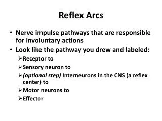
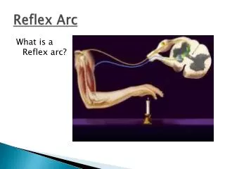
Reflex Arc • The reflex arc governs the operation of reflexes. Nerve impulses follow nerve pathways as they travel through the nervous system. The simplest of these pathways, which include only a few neurons, is called the reflex arc. Reflexes whose arc passes through the spinal cord are called spinal reflexes.
Parts of the Reflex Arc • Receptor – detects the stimulus. a) Description: the receptor end of a particular dendrite or a specialized receptor cell in a sensory organ. b) Function: sensitive to a specific type of internal or external change. • Sensory neuron – conveys the sensory info. to brain or spinal cord. a. Description: Dendrite, cell body, and axon of a sensory neuron. b. Function: transmit nerve impulses from the receptor into the brain or spinal cord.
Parts of the Reflex Arc 3. Interneuron: relay neurons. a. Description: dendrite, cell body, and axon of a neuron within the brain or spinal cord. b. Function: serves as processing center, conducts nerve impulses from the sensory neuron to a motor neuron. 4. Motor neuron: conduct motor output to the periphery. a. Description: Dendrite, cell body, and axon of a motor neuron. b. Function: transmits nerve impulse from the brain or spinal cord out to an effecter.
Parts of the Reflex Arc 5. Effector: a. Description: a muscle or gland. b. Function: Response to stimulation by the motor neuron and produces the reflex or behavioral action.
Spinal Reflexes Sensory feedback from muscle
Spinal Reflexes • The Myotatic Reflex • Stretch reflex: Muscle pulled tendency to pull back • Feedback loop • Discharge rate of sensory axons: Related to muscle length • Monosynaptic • Example: knee-jerk reflex
Spinal Reflexes The Myotatic Reflex
Spinal Reflexes • Gamma Motor Neurons • Muscle spindle • Intrafusal fibers: gamma • Extrafusal fibers: alpha • Gamma feedback loop provides more control
Spinal Reflexes • Reverse myotatic reflex • Regulate muscle tension • Golgi tendon organs
Spinal Reflexes Reciprocal inhibition Contraction of one muscle set accompanied by relaxation of antagonist muscle • Flexor reflex: Complex reflex arc used to withdraw limb from aversive stimulus • Crossed-extensor reflex: Activation of extensor muscles and inhibition of flexors on opposite side
Stretch and Deep Tendon Reflexes For skeletal muscles to perform normally: The Golgi tendon organs (proprioceptors) must constantly inform the brain as to the state of of the muscle. Stretch reflexes initiated by muscle spindles must maintain healthy muscle tone.
Muscle Spindles • Are composed of a few intrafusal muscle fibers that lack actin and myosin in their central regions, are noncontractile, and serve as receptive surfaces. • Muscle spindles are wrapped with two types of afferent endings: primary sensory endings of type Ia fibers and secondary sensory endings of type II fibers and secondary sensory endings of type II fibers. • These regions are innervated by gamma () efferent fibers. • Note: contractile muscle fibers are extrafusal fibers and are innervated by alpha () efferent fibers.
Operation of the Muscle Spindles • Stretching the muscles activates the muscle spindle. There is an increased rate of action potential in Ia fibers. • Contracting the muscle reduces tension on the muscle spindle. There is a decreased rate of action potential on Ia fibers
Operation of the Muscle Spindles
Stretch Reflex • Stretching the muscle activates the muscle spindle. • Excited motor neurons of the spindle cause the stretched muscle to contract. • Afferent impulses from the spindle result in inhibition of the antagonist inhibition of the antagonist. • Example: patellar reflex. Tapping the patellar tendon stretches the quadriceps and starts the reflex action. The quadriceps contract and the antagonistic hamstrings relax.
Golgi Tendon Reflex • The opposite of the stretch reflex. • Contracting the muscle activates the Golgi tendon organs. • Afferent Golgi tendon neurons are stimulated, neurons inhibit the contracting muscle, and the antagonistic muscle is activated. • As a result, the contracting muscle relaxes and the antagonist muscle contracts.
Monosynaptic Stretch Reflex Simplest reflex because it has only 1 synapse in the path of its arc. Muscle spindles contain the sensory receptors for the stretch reflex. Each spindle contains modified muscle fibers called spindle or intrafusal fibers (inside spindle), innervated by γ efferent fibers.
Monosynaptic Stretch Reflex The middle segment of each spindle fiber acts as a mechanical stretch receptor that is connected to a sensory afferent nerve to the spinal cord. Stretching of the muscle stretches the spindle fibers activating the muscle spindle stretch receptors and the associated sensory fibers.
The terminals of the spindle sensory fibers make direct excitatory synaptic contact with alpha motor neurons serving the ordinary muscle fiber (extrafusal fibers). • Contraction of these fibers shorten the muscle and relaxes the spindle fibers terminating the stretch reflex and muscle contraction.
Events in the Monosynaptic Stretch Reflex - Summary • 1. Passive stretch of a muscle (produced by tapping its tendon) stretches the spindle (intrafusal) fibers. • 2. Stretching of a spindle distorts its central (chain) region, which stimulates dendritic endings of sensory nerves. • 3. Action potentials are conducted by afferent (sensory) fibers into the spinal cord on the dorsal roots of spinal nerves. • 4. Axons of sensory neurons synapse with dendrites and cell bodies of somatic motor neurons located in the ventral horn gray matter of the spinal cord. • 5. Efferent impulses in the axons of somatic motor neurons (which form the ventral roots of the spinal nerves) are conducted to the ordinary (extrafusal) muscle fibers. These neurons are (alpha) motor neurons.
Events in the Monosynaptic Stretch Reflex - Summary • 6. Release of Ach from the endings of alpha motor neuron stimulates the contraction of extrafusal fibers, and thus the whole muscle. • 7. Contraction of the muscle relieves the stretch of its spindles, thus decreasing electrical activity in the afferent nerve fibers, and relaxes the spindle fiber and terminates the stretch reflex and muscle contraction. the stretch reflex and muscle contraction. • Note: By sending command to the motor neurons, the brain set a muscle’s length. The stretch reflex makes sure the muscle stay at that length. The stretch reflex is therefore important for maintaining muscle tone and upright posture.
The Deep Tendon Reflex • If you tap on the tendon of a muscle, it contracts. Its synergists contract and its antagonists are inhibited. - polysynaptic reflex. • A tap on the patellar tendon stretches the extensor muscle and its spindles.
The spindle discharges and excites the associated sensory fibers that excite the motor neurons to the extensor muscle. • - Contraction of the extensor muscle extends the lower leg (knee - jerk). • - Ipsilateral flexor muscle relax for extensors to function. • - Branches of the sensory fibers from muscle spindle activate inhibitory interneuron, which in turn, inhibit the motor neuron to the flexor muscle.
The Withdrawal Reflex (Flexor Reflex) • The automatic withdrawal of an extremity from a painful stimulus. • A polysynaptic reflex. • Sensory pain signals excite motor neurons to the flexor muscles, eliciting flexion and withdrawal of the leg. • Motor neurons to the extensor muscles are inhibited via inhibitory interneurons. • This would relax the extensors of the same leg.
The Crossed Extensor Reflex • A polysynaptic reflex. • E.g., Painful stimulation of one foot causes flexion (withdrawal) of the ipsilateral leg as well as the extension of the contralateral leg, to stabilize the posture; thus the ipsilateral leg flexors are activated and the extensors are inhibited and vice versa in the contralateral leg.
Superficial Reflexes • Biceps jerk reflex: the examiner places finger on the inside of the extended elbow over the tendon of the biceps muscle and the finger is tapped. The biceps contracts in response, and the forearm flexes at the elbow. • Triceps jerk reflex: tapping the short tendon of the triceps muscle close to its insertion near the tip of the elbow elicit this reflex. The muscle contracts in response, and the forearm extends, slightly. • Abdominal reflex: the examiner strokes the skin of the abdomen with a dull pin from the side of abdomen upward towards the midline and above the umbilicus. The umbilicus moves towards the stimulated region.
Superficial Reflexes • Ankle – jerk reflex (plantar reflex): tapping the Achilles tendon just above its insertion on the Calcaneous elicits this reflex. The response is plantar flexion, produced by contraction of the gastrocnemius and the solues muscles. • Cremastric reflex: this reflex is elicited in males by stroking the upper inside of the thigh. In response, the testis on the same side is elevated by contracting muscles.
Clinical Terms • Analgesia: loss or reduction in the ability to sense pain, without loss of consciousness. • Analgesic: pain – relieving drug. • Anesthesia: loss of feeling. • Ataxia: partial or complete inability to coordinate voluntary movements. • Epilepsy: Disorders of the CNS that is characterized by temporary disturbances in normal brain impulses; it may be accompanied by convulsive seizures and loss of consciousness. • Huntington disease: Hereditary disorders of the brain producing progressively worsening, uncontrollable dance-like movements and personality changes. • Neuralgia: sharp, recurring pain associated with a nerve, usually caused by inflammation or injury.