Microfluidic Device for High-Efficiency Single Cell Capture and Impedance Measurement
This study outlines the development of a microfluidic device for the precise capture of single HeLa cells, utilizing a flow rate of 5 ml/h and an infusion pump setup. The design includes three micro pillars to enhance cell capture probability, achieving around 10%. Impedance measurements were conducted to analyze cell behavior under varying frequencies and voltages, revealing significant decreases in impedance with increased operating voltage. The fabrication involved SU-8 molds and PDMS channels, ensuring optimal bonding and functionality. This device has potential applications in cell analysis and biomedical research.

Microfluidic Device for High-Efficiency Single Cell Capture and Impedance Measurement
E N D
Presentation Transcript
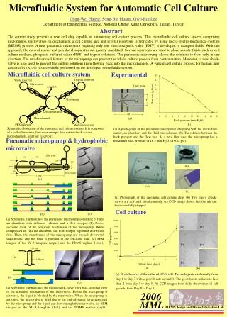
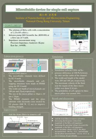
Microfluidic device for single cell capture 饒仁郁 余思緯 Institute of Nanotechnology and Microsystems Engineering, National Cheng Kung University, Tainan Conclusion • The solution of HeLa cells with a concentration • of 2.25×10⁵ cells/c.c • Infusion pump (KD Scientific Inc.,KDS100) at • a flow rate of 5 ml/h. • Impedance measurement using • Precision Impedance Analyzers (Kayne • Kerr Inc., 6440B). Fabrication process Simulation Simulation software:CFD–ACE+ pressure difference of 100 Pa between the inlet and the outlet of the channel. The width and length of the model were 200 and 100 μm, respectively, The maximum velocity of the fluid near the region of the three micro pillars was about 0.26 m/s. The probability of cell capture by three micro pillars was around 10%. Cr/Au=15/65 (nm) u The microfluidic channels were defined by SU-8 mold. The microfluidic channels and cell-trapping structures were made of PDMS using the SU-8 mold. The width and depth of microchannels are 100 μm and 25μm,respectively. The size of the micro pillars is 10μm x 10 μm x 25μm. The PDMS channels and the glass substrate with electrodes were treated by O2 plasma (800 W, 12 sec) to improve bonding strength. Result and discussion • Add the front filters to remove call debris • Increasing the frequency reduced the magnitude of the HeLa cell impedance • Increasing the operating voltage decreased the magnitude of the HeLa cell impedance. • At an operating voltage of 0.8 V, the phase decreased from −66.9 to −72.2° below30 kHz but the phase increased from −72.2 to −67.5° at 0.8 V between 30 and 100 kHz Improvement • Add the front filters to remove call debris