Download
1 / 1
10 likes | 141 Vues
This study delves into the microbial communities thriving on wrestling mats, showcasing the resilience of bacteria despite cleaning efforts. By isolating colonies, conducting gram staining, and live/dead staining, the experiment reveals bacteria types present and their survival. The findings underscore the importance of effective disinfection to prevent skin infections among athletes.

E N D
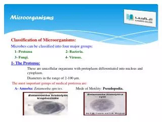
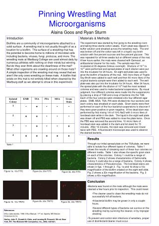
Figure 1a. 10x FITC Figure 2a. 20x FITC Figure 3a. 40x FITC Figure 2b. 20x TRITC Figure 1b. 10x TRITC Figure 3b. 40x TRITC Introduction Biofilms are a community of microorganisms attached to a solid surface. A wrestling mat is not usually thought of as a location for a biofilm. The surface of a wrestling mat has the potential to become home to millions of microbial cells including bacteria, viruses, fungi, protozoa, and more. The wrestling mats at Wartburg College are used almost daily by numerous athletes with nothing on their minds but winning. But do they ever think about the cleanliness of the mat? What other organisms are crawling around on those mats? A simple inspection of the wresting mat may reveal that we aren’t the only ones wrestling on these mats. A biofilm that exists on this mat is not entirely killed when cleaned by the Wartburg staff as we attempt to show in this experiment. Materials & Methods The experiment was started by first going to the wrestling room and taking three sterile cotton swabs. Each swab was dipped in buffer solution and streaked across the wrestling mats. The end was broken off and the cotton was left in a tryptic soy broth solution with five percent sucrose and five percent glucose. This was repeated for all three swabs but on different areas of the mat. A few hours earlier, the mats were cleaned with Gemesol, an antibacterial cleaner for the mats. The sample was then incubated at 35 degrees Celsius overnight. Dilutions of 10-2 to 10-9 were performed and spread plates of each dilution were put into the incubator for 24 hours. An eight well plate was used to grow the biofilm of bacteria off the mat. 400 micro liters of Tryptic Soy Broth were added to each well and then 50 micro liters of the original bacteria sample were then added to each well. The well plate was also put into the incubator for 24 hours. After 24 hours, the spread plate with the dilution of 10-6 had the most isolated colonies and was used to make bacterial suspensions. By visual judgment, four different colonies were made into the suspensions by placing a drop of TSB and a loop of bacteria into the TSB. Each of the four colonies were streaked onto four different agar plates. EMB, MSA, TSA, PIA were divided into four sections and each colony was streaked on each plate. Gram stains were then performed on each of the four bacterial suspensions to discover if they were gram positive or gram negative. A live dead stain was performed by first mixing 1ml of 10% NaCl with 3 micro liters of live/dead stain while in the dark. The liquid in the eight well plate was drawn off and PBS was added to rinse the plate twice. Once the PBS was removed the second time, 10 micro liters of live/dead stain was added in the dark and let stand for 15 minutes. After 15 minutes, the stain was removed and rinsed twice with PBS. A fluorescent microscope was used to observe the stained bacteria. Pinning Wrestling Mat Microorganisms Alaina Goos and Ryan Sturm Results Through our initial spread plate on the TSA plate, we were able to isolate four different types of colonies. Table 1 shows the results of streaking each of these four colonies on different media. Table 1 also shows the specific gram stain on each of the colonies. Colony 1 could be a range of bacteria. Colony 2 shows characteristics of Salmonella. Colony 3 could also be a range of bacteria. Colony 4 shows characteristics of Pseudomonas. The live/dead stains are shown in figs. 1 through 3. Fig. 1a and 1b show a 10x magnification of the biofilm created on the eight well slide. Fig. 2 shows a 20x magnification of the bacteria. Fig. 3 shows a 40x magnification. Table 1 • Conclusion • Bacteria was found on the mats although the mats were cleaned a few hours prior to inspection. This could mean: • The cleaner used to clean the wrestling mats do not successfully kill all bacteria. • A bacterial biofilm may be grown in only a couple hours. • Several different types of bacteria can survive on the wrestling mat by surviving the cleaner, or by improper cleaning • To prevent and control skin infections of wrestlers, proper use of disinfectant/cleaner must occur. References Difco Laboratories. 1998. Difco Manual. 11th ed. Sparks, MD Becton Dickenson & Co. Harley, John P., Donald A. Klein, and Lansing M. Prescott. 5th ed. New York, NY: The McGraw-Hill Companies, Inc., 2002. 620-621.