Download
1 / 87
870 likes | 908 Vues
Learn about common congenital urinary tract abnormalities and benign prostatic hypertrophy, including symptoms, diagnosis, treatment, and surgical options. Explore conditions like urethral stricture, renal agenesis, and more.

E N D
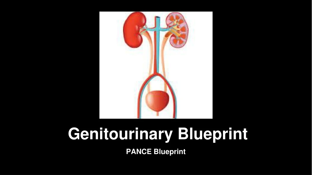
Genitourinary Blueprint PANCE Blueprint
GU Tract Conditions • Benign Prostatic Hyperplasia (BPH) • Benign Prostatic Hypertrophy (BPH) presents with polyuria, nocturia, hesitancy, urgency and weak urinary stream. • Symptoms of BPH appear gradually over a number of years • Incidence increase with age • BPH is not believed to be a risk factor for prostate cancer
Benign Prostatic Hypertrophy • Differential diagnosis includes: urethral stricture, bladder neck contracture, carcinoma of the prostate, carcinoma of the bladder, bladder calculi, urinary tract infection, prostatitis, and neurogenic bladder • Recommended test is urinalysis and serum PSA and renal panel • Patients with symptoms should have a digital rectal exam to assess prostate size and detect nodules and malignancy • Optional tests include maximal urinary flow rate (normal > 15 mL/sec), post residual urine volume, and urine cytology • Many agents used for treatment of BPH • Alpha 1 Adrenergic Antagonists (Terazosin, Doxazosin, and Tamsulosin) act against the dynamic component of bladder outlet obstruction by relaxing smooth muscle in the bladder neck, prostate capsule, and the prostatic urethra
Benign Prostatic Hypertrophy • -5 Alpha Reductase Inhibitors (Finasteride and Dutasteride) act by reducing the size of the prostate gland • Patients with frequency, urgency, and incontinence may be due to an overactive bladder • Surgical treatment for BPH includes TURP (transurethral resection of the prostate) • Open Prostatectomy can be for those who fail TURP • Urethral stent may be effective for selected patients
Congenital Abnormalities • Renal Agenesis- is almost always unilateral and does not cause symptoms. It is when the kidney is absent or underdeveloped • Renal Hypoplasia-this appears as one small kidney with the other one larger than normal. Small kidneys have small arteries that can lead to hypertension. A nephrectomy maybe needed to correct this. • Supernumerary Kidneys-third kidney is very rare and not be confused with the relatively common unilateral duplication of the renal pelvis
Congenital Abnormalities • Renal Dysplasia and Multicystic Kidney- multicystic kidney is usually only seen in one kidneys and is an irregularly lobulated mass of cysts and usually absent or atretic ureter. Usually associated with other abnormalities such as a ureteropelvic junction obstruction. Dysplasia of the renal parenchyma is seen with urethral obstruction or reflux present in early pregnancy or obstructed ureter • Simple Renal Cyst-can be inherited or acquired. Significant renal damage is rare but usually requires only continuous followup. • Renal Fusion-the most frequent abnormality seen is a horseshoe kidney containing two excretory system and two ureters • Ectopic Kidney-with a simple ectopic kidney the kidney does not ascend properly and is found in the pelvis or over the brim. It is prone to infection and obstruction. Less common is crossed ectopy without fusion.
Congenital Abnormalities • Ureteral Atresia-the ureter may be absent or fail to extend to the bladder. It is associated with ipsilateral absent or multicystic kidney. Bilateral atresia is incompatible with life • Duplication of ureter-one of the most common congenital malformations. It is often asymptomatic but may have recurrent UTI's • Ureterocele- This is a sacculation of the bladder end of the ureter that can occur either in the bladder or ectopically. It may be asymptomatic or causes obstruction, incontinence or infection. • Ectopic Urethral Orifice-usually occurs with ureterocele and duplication of the ureter but single ectopic ureters are seen. Males may get epididymitis because ureter drains into the vas deferens • Obstructed Mega Ureter-caused by obstruction at the UVJ
Congenital Abnormalities • Bladder Exstrophy- The absence of the anterior wall of the bladder, with the ureters delivering urine into the lower abdomen. Requires surgical correction • Persistent urachus- This appears as a draining umbilical sinus and can become infected • Contraction of the Bladder Neck-common cause of reflux, bladder diverticulae or irritable bladder • Apenia -absence of a penis. Rare.
Congenital Abnormalities • Megalopenis- the penis enlarges rapidly in childhood due to high levels of production of testosterone • Micropenis- Small but otherwise normally formed penis with a stretched length of less than 2.5 standard deviation below the mean. • Urethral Stricture-severe case can result in damage to the bladder and hydronephrosis due to back pressure of the urine • Hypospadias-urethral meatus found on the underside of the penis. • Epispadias- this is a defect of the dorsal wall of the urethra. The urethra is displaced dorsally so that it opens on the top of the penis in males
Congenital Abnormalities • Labial Fusion-fused labia minora causing recurrent infections as a result of obstruction to urinary flow • Clitoral Hypertrophy-this is caused by exposure to fetal androgens • Polycystic Kidney Disease-the most common cause of serious renal inherited disease. It is an autosomal dominant condition. • Medullary Sponge Kidney-can affect one or both kidneys. There are ectatic and cystic changes of the medullary and papillary collecting ducts. • Potter's Syndrome-describes the typical physical appearance caused by pressure in utero due to oligohydraminos due to bilateral renal agenesis
Cryptorchidism • Most cyrptorchid testes are undescended but some are absent due to agenesis or atrophy • True undescended testes have stopped short along their normal path of descent into the scrotum • Cryptorchism is defined as testis not in the scrotum not descended into the scrotum by four months of age • In most instances referral is appropriate if testicles not descended between 6-12 months of age
Cryptorchidism • Complications of undescended testicles include inguinal hernia, testicular torsion, testicular trauma, subfertility, and testicular cancer. • Surgery is recommended ASAP after the child is 6 months of age and should definitely be completed before 2 years of age • Orchioplexy is the procedure of choice for repositioning palpable undescended testicles • Exploratory surgery is recommended for non palpable undescended testis
Erectile Dysfunction • Erectile Dysfunction (ED) is defined as the consistent or recurrent inability to acquire or sustain an erection of sufficient rigidity and duration for sexual intercourse • Risk factors for ED include: obesity, smoking, watching television, dyslipidemia, obstructive sleep apnea, prescription meds (SSRI's spironolactone, clonidine, methyldopa, thiazide diuretics, cimetidine, and ketoconazole) and cardiovascular disease
Erectile Dysfunction • -Other factors may lead to ED such as psychosocial factors (depression and stress), neurological (spinal cord injuries, MS, or dementia), bicycling, testosterone deficiency, hyperthyroidism, hyperprolactinemia, and hypothyroidism • Normal erections require blood flow from the hypogastric arterial system into erectile chambers. High levels of intrapenile nitric oxide act as a local neurotransmitter to facilitate the relaxation and cause erection • Treatment includes Phosphodiesterase-5 Inhibitors (PDE-5)-including sildenafil, vardenafil, and tadalafil • These all act to increase intracavernosal cyclic GMP levels, and each one has been proven to be effective in restoring erectile dysfunction. • There is intrapenile injection options with PDE-5 inhibitors as well as vacuum devices available if these other methods are not an option.
Hydrocele/Varicocele • Hydrocele is a collection of peritoneal fluid between the parietal and visceral layers of the tunica vaginalis, the investing layer that directly surrounds the testis and the spermatic cord • Hydroceles are believe to arise from an imbalance of the secretion and reabsorption of fluid form the tunica vaginalis • Symptom of hydrocele of pain and disability and increasing size of the mass • Hydroceles can be from epididymitis, torsion, appendiceal torsion, and can be idiopathic • Ultrasound can be used when the diagnosis is in question
Hydrocele/Varicocele • Surgical excision is the most common treatment of hydrocele • Varicocele is caused by a dilation of the pampinform plexus of the spermatic veins. • Varicocele occurs in the left hemiscrotum usually • Symptoms of varicocele include dull, aching left scrotal pain, typically worse when standing better when setting • Varicocele can cause decreased fertility • Treatment of varicocele surgical treatment ligating the gonadal vein so that retrograde blood flow can no longer reach the plexus of veins in the scrotum.
Urinary Incontinence • Urinary incontinence is the involuntary leaking of urine • Four main types of urinary incontinence are urge, stress, mixed, and overflow incontinence • The types of incontinence overlap • Cause of urinary incontinence: UTI, those that originate from the urinary tract, those caused by a systemic condition (diabetes mellitus), and those from a reversible cause (medications)
Urinary Incontinence • Urgency incontinence have symptoms of incontinence accompanied by urgency. Partly caused by detrusor overactivity • Stress incontinence have involuntary leakage that occurs with effort, sneezing, coughing laughing, and anytime there is an increase in intra-abdominal pressure • Overflow incontinence is involuntary, continuous leakage or dribbling and incomplete bladder emptying. Caused by impaired detrusor contractility or bladder outlet obstruction • Look for potentially reversible causes • Need bladder stress test, post void residual volume and urodynamic testing
Urinary Incontinence • Surgery may be helpful for stress incontinence and other instances • Weight loss can decrease the episodes of stress incontinence • Bladder retraining can help with urgency, stress, or mixed incontinence • Kegel exercises are effective for urgency, stress, and mixed incontinence • Continence pessaries may be used for stress incontinence • Antimuscarinics such as darfenacin, fesoterodine, oxybutin, solifenacin, and tolterodine are effective for urgency and mixed incontinence or overactive bladder • Other modalities include electrical stimulation and magnets • Surgery is a last resort for intractable urgency incontinence
Nephrolithiasis/Urolithiasis • Several types of kidney stones: calcium oxalate, calcium phosphate, uric acid, cysteine and struvite stones • Calcium Oxalate stones are the most common kidney stones. Risk factors for development include low urine volume, higher urine calcium excretion, higher urine oxalate excretion, and lower urine citrate excretion • Calcium phosphate stones make up about 15 percent of stones and can be seen in combination with calcium oxalate or struvite stones. Risk factors include lower urine volume, higher urine calcium excretion, lower urine citrate excretion, higher urinary pH, and higher urine phosphate excretion.
Nephrolithiasis/Urolithiasis • Uric acid stones are radiolucent. Risk factors include lower urine volume, high urine uric acid excretion, and lower urine pH • Struvite stones only form in the presence of urease producing bacteria (proteus). High urine pH and infection are risk factors for development of these struvite stones. • Presenting symptoms typically include flank pain radiating to lower abdomen, nausea, vomiting, dysuria, hematuria, and polyuria. May have fever and chills accompanying it.
Nephrolithiasis/Urolithiasis • Ninety percent of stones less than 4 mm will pass with a couple weeks. • Kidney stones greater than 5 mm is associated with decrease in spontaneous passage rate • Initial workup should include CBC, BMP, and Urinalysis. Imaging studies CT scan without contrast is ideal, but if radiation exposure is a concern renal ultrasound can demonstrate hydronephrosis and shadows which can indicate the presence of stones. • Analgesics Toradol is ideal if not pregnant, bleeding is not a concern, and renal function is intact. Morphine and Dilaudid can be used as adjuncts. Zofran or Phenergan for nausea. • Flomax 0.4 mg daily has been shown to relax ureter and help with spontaneous passage of stone
Nephrolithiasis/Urolithiasis • If stones are septic and obstructing emergent decompression of the collecting system is needed with percutaneous drainage or ureteral stenting with antibiotics (cipro or rocephin) • Surgical therapy for stones that do not or cannot pass spontaneously include lithotripsy, stenting, or basket retrieval
Paraphimosis/Phimosis • Paraphimosis-refers to retracted foreskin in an uncircumcised or partially circumcised glans penis that cannot be returned to its normal position • Phimosis-a tight foreskin that cannot be retracted to expose the glans penis. Can be normal or physiologic in young children. Older adults may get infections such as balanoposithis or inflammatory scarring
Paraphimosis/Phimosis • Paraphimosis can cause impairment of venous and lymphatic flow from the constricting ring and cause engorgement of the glans penis and swelling. The arterial flow becomes compromised over hours to days. Penile necrosis, infarction of the glans, or gangrene can occur • Treatment consists of timely reduction of the foreskin over the glans penis and requires pain control
Testicular Torsion • Testicular torsion is a urologic emergency that is most common in neonates and post pubertal boys • Testicular torsion can occur at any age • Testicular torsion results from the inadequate fixation of the testis to the tunica vaginalis. The testis may twist on the spermatic cord causing ischemia from reduced arterial blood flow and venous obstruction • Testicular torsion may be from trauma or can occur spontaneously. Can also reduce spontaneously and be occur intermittently
Testicular Torsion • It is generally felt that testis suffered irreversible damage after twelve hours of torsion. • Clinically there is usually sudden onset of pain occurs usually after several hours of vigorous activity or minor trauma. There can be nausea and vomiting. • Classic exam finding include a asymmetric high riding testis on the affected side with the long axis of the testis oriented transversely instead of longitudinally secondary to shortening of the spermatic cord. This is called a Bell Clapper deformity
Testicular Torsion • Diagnosis is made by ultrasound • Treatment is surgery and involves detorsion and fixation • If surgery is over two hours from happening attempts at manual detorsion should be attempted by rotating outward toward the thigh.
Cystitis • Acute cystitis refers to infection of the bladder (lower urinary tract) • The pathogenesis of UTI is colonization of the bacteria by uropathogens usually from fecal flora (in women) in the urethra and they ascend up through the lower urinary tract • Pyelonephritis is when the bacteria ascend up the ureter to the kidney • Uncomplicated UTI in women consists mainly of Escherichia Coli (75-95%) with other bacteria making up the rest (enterobacter, Proteus Mirabillis, and Klebsiella pneumoniae)
Cystitis • Among non pregnant women, colonization of organisms such as lactobacilli, enterococci, and group B strep from clean catch urine most commonly represents contaminated urine. If these are in symptomatic women in high counts they should be treated as a UTI • Antibiotic resistance > 20 percent with ampicillin and bactrim, less than 10 percent with fluoroquinolones • Augmentin and first generation cephalosporins are less than 10 percent usually • Nitrofurantoin can also be used for pregnant patients • Symptoms include dysuria, frequency, urgency, suprapubic pain and hematuria • Urinalysis for the evaluation of pyuria is the most important test for a UTI
Cystitis • WBC with cast are diagnostic of an upper urinary tract infection • Leukocyte esterase (>10 leukocytes per high power field) sensitivity 75-96 sensitivity • Treatment usually for up to 7 days with uncomplicated UTI • Asymptomatic pyuria in men is rare. Should be done after a TURP • Risk factor for these infections are anal intercourse and the lack of circumcision
Cystitis • A complicated UTI can be upper or lower urinary tract infection but is associated with a risk factor of failing therapy. Some of these risk factors include: diabetes, pregnancy, symptoms more than 7 days, renal failure, nosocomial infection, urinary tract obstruction, instrumentation, renal transplant, and immunosuppression • Pyelonephritis is an infection of the kidney characterized by fever, flank pain, nausea, and vomiting. Symptoms of a lower urinary tract infection are usually present
Epididymitis • Epididymitis is the most common infection seen in the outpatient setting for scrotal pain. • Epididymitis is an infection of the epididymis that lays superior to the testis • More advanced cases present with testicular swelling and pain epididymorchitis • Acute epididymitis is less than 6 weeks • Chronic epididymitis is more than 6 weeks
Epididymitis • Physical exam shows tenderness to the epididymis with or without swelling. • Urinalysis should be obtained but the urine is usually negative especially in patients without urinary complaints • Ultrasound should be performed to assess for the presence of torsion • All men should be treated empirically with regimens that cover C. Trachomatis and N. Gonorrhoeae (rocephin 250 mg IM plus Doxycyline 100 mg BID for 10 days) • Men that are over 35 that practice anal intercourse should be covered with rocephin plus ofloxacin or levaquin for 10 days • Epididymitis that is non infectious is rare and caused by trauma, vasculitis or autoimmune disorders
Orchitis • Orchitis is inflammation or infection of the testis • Orchitis without epididymitis is extremely rare • Mumps should be considered as a diagnosis with isolated orchitis • In immunized adults, viruses similar to those in children can cause orchitis • When seen concurrently with epididymitis should be treated with the same antibiotic coverage as isolated epididymitis
Prostatitis • Acute bacterial prostatitis is an infection of the prostate usually caused by gram negative organisms • Entry of the microorganisms into the prostate almost always occurs via the urethra • There may be an infection of the epididymis at the same time • Prostatitis can occur in the presence of cystitis, urethritis, or other urogenital infections • Common causative organisms of prostatitis include E.Coli, Proteus, Klebsiella, Enterobacter, Staphylococcus aureus, and Pseudomonas
Prostatitis • If patient is less than 35, the patient should be treated for gonorrhea and chlamydia • Diagnosis is made digital rectal exam • Treatment of acute prostatitis can include bactrim, cipro, levaquin for 6 weeks • Chronic prostatitis is recurrent urogenital symptoms with evidence of bacterial infection of the prostate • Pathogenesis of chronic prostatitis is the same as acute prostatitis • Common organisms of chronic prostatitis is enterococcus, klebsiella, proteus, and pseudomonas. Chlaymida has been an associated organism • For chronic prostatitis fluoroquinolones or bactrim is needed for at least 6 weeks
Pyelonephritis • Pyelonephritis is an infection of the kidney • Pyelonephritis presents with fever, chills, flank pain, nausea, vomiting, dysuria, polyuria, and/or hematuria • Common organisms include E.Coli, Proteus, Klebsiella, and Staphylococcus • Treatment should involve cipro or levaquin, cephalosporins, or beta lactam inhibitors • Antibiotics should be administered for 10-14 days
Urethritis • Urethritis is inflammation of the urethra is a common manifestation of sexual transmitted diseases • N. Gonorrhea is the most common cause of urethritis in the United States • C. Trachomatis is the most common cause of non gonococcal urethritis • Clinical manifestations of urethritis include dysuria, and penile discharge from the urethral meatus
Urethritis • Diagnosis is made by penile discharge on examination, positive urethral swab, positive leukocyte esterase on first catch urine, or positive urine culture • Gonorrhea is treated with 250 mg of rocephin • Chlamydia is treated with 1 gram of Zithromax or 100 mg of doxycycline BID for 10 days
Bladder Carcinoma • Bladder carcinoma is the most common malignancy of the genitourinary system • Transitional cell carcinoma is the most most common histologic type • Bladder cancer ranges from superficial (not involving the muscle), to muscle invasive, and metastatic disease • Bladder cancer typically presents with painless hematuria • Bladder cancer can have frequency, urgency and dysuria
Bladder Carcinoma • Pain associated with bladder cancer is usually associated with locally advanced metastatic tumor • Physical exam is usually unremarkable but may feel pelvic mass in patients with advanced metastatic disease • Workup should include cystoscopy, urinalysis, and urine cytology. CT scan of abdomen and pelvis to evaluate the collecting system and the renal cortex • Tumors are staged with TMN system • Bladder cancer can be treated with surgery and/or chemotherapy
Prostate Carcinoma • Prostate cancer is the second most common cancer in men • The clinical spectrum of prostate cancer ranges from microscopic, well differentiated tumor to higher grade cancer that causes metastasis • Most men undergo diagnosed with prostate cancer undergo a biopsy because of an abnormal PSA • PSA levels can also be elevated with BPH and Prostatitis • On digital rectal exam, asymmetric areas of induration or frank nodules are indicated for prostate cancer and a prostate biopsy should be performed regardless of PSA
Prostate Carcinoma • Transrectal ultrasound (TRUS) is used to evaluated abnormalities detected by digital rectal exam and guide to sites of biopsies • Patients may have no symptoms of prostate cancer in the early stages • Symptoms if present may include: urinary frequency, urgency, nocturia and hesitancy. Hematuria and hematospermia are common in older men • Bone pain may be present with metastatic disease • Patients with low risk clinically localized prostate cancer external beam radiation therapy and radical prostatectomy
Prostate Carcinoma • Patents with intermediate risk standard of treatment includes radiation therapy, radical prostatectomy with pelvic lymph node dissection • Patients with high risk standard of treatment includes radiation therapy with radical prostatectomy • In some instances, castration and chemotherapy is need
Renal Cell Carcinoma • Renal cell carcinomas originate from the renal cortex and make up the majority of renal neoplasms. • Renal Cell Carcinoma patients present with flank pain, hematuria, and a palpable abdominal mass (small percentage of patients). Most patients have no symptoms • Advanced disease may have inferior vena cava involvement causing lower extremity edema, ascites, and hepatic dysfunction
Renal Cell Carcinoma • May have associated anemia, hepatic dysfunction fever, and hypercalcemia with advanced disease • Diagnosis is made with renal ultrasound or CT scan of Abdomen and Pelvis • MRI of the abdomen is helpful when the CT scan or ultrasound is inconclusive or IV contrast allergy • Staging with the TMN staging system
Renal Cell Carcinoma • Surgery is considered curative for those who do not have distant metastasis • Molecular Therapy is used as an adjunct for those with advanced RCC. Cytokine therapy is also used. • Chemotherapy is used in some instances