Download
1 / 18
180 likes | 383 Vues
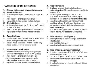
‘In families with mitochondrial disorders, different family members can present with different clinical manifestations’. Discuss this statement with respect to the principles defining mitochondrial genetics. Patterns of Inheritance Session 4 th September 2009 Kirsten McKay.

E N D
‘In families with mitochondrial disorders, different family members can present with different clinical manifestations’. Discuss this statement with respect to the principles defining mitochondrial genetics. Patterns of Inheritance Session 4th September 2009 Kirsten McKay
Mitochondrial genome • The main role of the mitochondria is in aerobic respiration including the tricarboxylic acid cycle and oxidative phosphorylation to produce ATP • Circular dsDNA molecule of 16,569 base pairs • 93% coding • Polycistronic transcription • Modified genetic code • 37 genes; 22 tRNAs, 2 rRNAs and 13 polypeptides (all OxPhos) • 10x more prone to errors • 2-10 mtDNA molecules per mitochondrion • 1,000-10,000 mitochondria per cell
mtDNA inheritance • mtDNA is maternally inherited • Cells are polyploidy • HOMOPLASMY = no detectable variation in mtDNA molecules of an cell/tissue/individual • HETEROPLASMY= detectable variation at one or more loci in mtDNA within one cell/tissue/individual • MUTATIONAL LOAD • Heteroplasmic mutations can be transmitted at various levels due to the BOTTLENECK during oocyte maturation
Specific mutations - LHON • Leber’s Hereditary Optic Neuropathy (LHON) – frequency ~1/30,000 most common mt disease • Bilateral loss of vision caused by degeneration of the optic nerve. • Susceptibility of retinal ganglion cells • Specific mutations in genes that encode for components of Complex I of the oxidative phosphorylation system. • Three most common primary mutations; • G11778A in ND4 gene (69%, most severe) • G3460A in ND1 gene (13%, less severe) • T14484C in ND6 gene (14%, least severe) • Other 5% are considered secondary mutations • Found in one individual or family • Found in association with primary mutations
Specific mutations - LHON • Penetrance is incomplete • 50-60% in males and 10-20% in females • Mutational load, tissue distribution and threshold effect; • 10-15% heteroplasmic • Levels of mutant mtDNA in peripheral blood are not always representative of levels in optic nerve • Patients are either affected or unaffected and it is likely that a threshold level of ~60% mutant mtDNA is required. • Modifying factors; • mtDNA background - haplogroups • Nuclear genes – likely to be X-linked • Environmental factors – ?alcohol, tobacco • General guidelines; • More mutant mtDNA; more likely to be penetrant! • More mutant mtDNA; more likely to be transmitted! • BUT not always the case!
Specific mutations – m.8993T>G/C • MILS=maternally-inherited Leigh syndrome • Progressive neurologic disease with motor and intellectual developmental delay • Brainstem and/or basal ganglia disease • Characteristic neuropathological features • Onset 3-12 months, death by 2-3 years • Various mtDNA genes including ATP6 • NARP=Neurogenic muscle weakness, Ataxia and Retinitis Pigmentosa • Peripheral neuropathy • Cerebral and cerebellar atrophy • Variable ocular manifestations • Later childhood or adult onset • Mutations in ATP6 gene • Intermediate phenotypes • Any combination of NARP and MILS • Other symptoms of mtDNA disease
Specific mutations – m.8993T>G/C • Common mutation in ATPase subunit 6 gene (complex V) • m.8993T>G or m.8993T>C • Minimal tissue-dependent and age-dependent variation • Phenotype is more predictable based on mutation load; • Up to 60% - unaffected • 60-90% - NARP • >90% - MILS • MILS/NARP are multi-system disorders and show a continuum of phenotypes dependent upon Mutational Load!
Specific mutations – m.3243A>G • MELAS – mitochondrial encephalo-myopathy syndrome with lactic acidosis and cerebro-vascular accident episodes • Maternally inherited diabetes with deafness • Maternally inherited cardiomyopathy • CPEO– chronic progressive external opthalmoplegia • Very diverse phenotypes including disorders of the brain, ears, eyes, skeletal muscle, cardiac muscle, and metabolism.
Specific mutations – m.3243A>G • MT-TL1 gene - tRNA Leucine • Mutational load- variable within families • Tissue distribution– muscle in CPEO, nerves in MELAS • Threshold effect– certain tissues may be more susceptible to mitochondrial dysfunction than others • Modifying factors– mitochondrial, nuclear, environmental • Progression – sequential muscle biopsies show increased levels of mutant mtDNA
mtDNA deletion syndromes • 3 overlapping phenotypes- may be present in different family members or progress in one individual over time • Kearns-Sayre syndrome (KSS)– multisystem disorder, childhood onset, retinopathy and opthalmoplegia • Pearson syndrome– sideroblastic anaemia, exocrine pancreas dysfunction, death in infancy • CPEO– variable myopathy, ptosis and opthalmoplegia • No specific region of mtDNA involved; at least one tRNA gene is deleted • Usually flanked by short repeat sequences • Usually de novo, rarely inherited • Common deletion 4977 base pairs
mtDNA deletion syndromes • Phenotype is characterised by; • Mutational load– threshold effect is generally 80-90% • Tissue distribution– KSS in all tissues, PS in haematopoietic cells and CPEO in skeletal muscle • Mitotic segregation– mutational load in dividing blood cells decreases with age and increases in post-mitotic cells therefore PS can progress to KSS
Nuclear DNA mutations • Deficiency of ETC • AR Leigh syndrome • Deficiency of mtDNA maintenance • AD CPEO • POLG1 • mtDNA depletion syndrome • DGUOK • SUCLA2
Associations with common diseases • Ageing • Age-related damage is caused by ROS • mtDNA mutations accumulate in ageing tissue – point mutations and deletions • Each mutation has ML of less than 1% but combined can impair OXPHOS leading to more ROS • Mouse model shows reduced lifespan, hair loss, osteoporosis, reduced fertility, reduced activity and weight loss. • Cancer • Cancer cells acquire mtDNA mutations – solid tumours and leukaemias • Many are homoplasmic – mechanism unclear • Pathogenicity is also unclear
Associations with common diseases • Parkinson’s disease • Common neurodegenerative disease • Risk factors include family history + environmental factors • Mostly sporadic however several familial forms have been identified • Some encode proteins that function in the mitochondria eg PINK1 • Mitochondrial dysfunction leads to neuronal death. • mtDNA variants may modify phenotype in PINK1 PD • In addition, mtDNA inheritance often appears to be sporadic, therefore direct involvement of mtDNA in predisposition to PD is very possible
Family planning options • Prenatal diagnosis is difficult • Heteroplasmy and tissue distribution • Bottleneck phenomenon • Poor correlation between ML and disease severity. • Women with mtDNA mutations have limited options • CVS + PGD m.8993T>G/C • Oocyte donation • Oocyte sampling • Superovulation is stimulated and then all oocytes collected and analysed for mtDNA mutation • Oocytes not used for implantation, but as a sample of oocyte population to help predict outcome of pregnancy. • Cytoplasmic or nuclear transfer
Family planning options • Nuclear transfer
References • Strachan & Read 3rd Edition • Gene Reviews – Mitochondrial Disease Overview • http://www.mitomap.org/ • OMIM • ‘Mitochondrial DNA Mutations in Human Disease’ Taylor & Turnbull Nature Reviews Genetics 2005 vol6 389-402 • ‘Genetic diseases of human mitochondrial DNA’ Solano et al Salud Publica Mex 2001;43:151-161 • ‘Inherited mitochondrial optic neuropathies’ Yu-Wai-Man et al J Med Genet 2009;46:145–158 • ‘Leber hereditary optic neuropathy - a disease with a known molecular basis but a mysterious mechanism of pathology’ Mroczek-Tonska et al J. Appl. Genet. 44(4), 2003, pp. 529-538 • ‘Segregation of mitochondrial DNA (mtDNA) in human oocytes and in animal models of mtDNA disease: clinical implications’ Poulton & Marchington Reproduction 2002 123 p751-755 • ‘The transmission of OXPHOS disease and methods to prevent this’ Jacobs et al Human Reproduction Update 2006 vol 2 p119-136 • ‘PINK1-associated Parkinson’s disease is caused by neuronal vulnerability to Calcium-Induced Cell Death’ Gandhi et al Molecular Cell 2009 33(5-3) 627-638