Download
1 / 72
750 likes | 1.28k Vues
Marine Invertebrates: Phylum Porifera. Phylum Porifera. Porifera – “ pore bearers ” Representative Organisms: Sponges. Intro. Characteristics of Porifera: 1) No definite symmetry . 2) Body multicellular , few tissues, no organs .

E N D
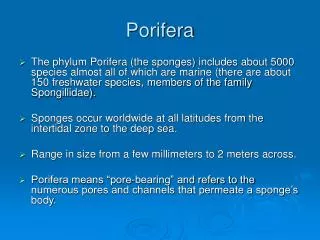
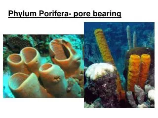
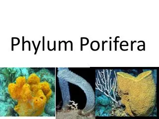
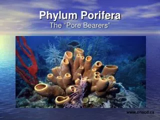
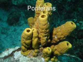
Phylum Porifera • Porifera – “pore bearers” • Representative Organisms: • Sponges Intro
Characteristics of Porifera: 1) No definite symmetry. 2) Body multicellular, few tissues, no organs. 3) Cells and tissues surround a water filled space but there is no true body cavity. 4) All are sessile (part of the benthic population) 5) Reproduce sexually or asexually. 6) No nervous system. - Have myocytes near ostia and osculum (contract to prevent water from entering (defense)7) All are filter feeders. 8) Skeleton of spicules.
Introduction • Sponges • Groups of sedentary organisms once thought to be plants. • Most primitive of the multicellular animals. • Simple body organization having a system of water canals. • More than 5,000 species of marine sponges and a few freshwater species. • Was once a profitable industry, however overfishing, disease and pollution have taken their toll. • Synthetic sponges have replaced the natural product.
Morphology path of water • Three Layers: • Ectoderm – Mesohyl – Endoderm • PINOCOCYTES: flattened layer of epithelial cells on the external surface. • OSTIA: openings in the ectoderm (surrounded by pore cells) • CHOANOCYTES (collar cells): makes up the feeding chamber; have flagella which creates currents
Morphology, cont’d • AMEBOCYTES: wandering cells • located in the mesohyl • Secrete spicules • Transport & store food particles • Transform themselves to repair sponge • SPICULES: give support ; made of calcium carbonate or silica; important in classification • SPONGIN: elastic framework made of protein fiber • OSCULUM: opening at top where water and wastes exit
Feeding Strategy: Suspension feeding http://www.youtube.com/watch?v=tl_gjeXKDps&NR=1 • Filter feeders – • Water is actively pumped through ostia into feeding chamber • Feeding chamber is composed of collar cells (choanocytes) which have flagella and collar to trap food • Food is digested in vacuoles (intracellular) • Filtered water then leaves through osculum (opening)
reproduction • Reproduction • Asexually • Budding • Regeneration • Sexually through broadcast spawning • Most are hermaphrodites (can produce both male and female gametes at different times) • Release of male gametes into water • Fertilization occurs internally
Tube Sponge Image Source: http://www.pbs.org/kcet/shapeoflife/animals/porifera3.html
Indonesian Sponge Image Source: http://www.pbs.org/kcet/shapeoflife/animals/porifera5.html
Examples of Sponge Spicules Image Source: http://www.pbs.org/kcet/shapeoflife/animals/porifera4.html
Branching Sponge Image Source: http://www.pbs.org/kcet/shapeoflife/animals/porifera2.html
Red Barrel Sponge Image Source: http://www.pbs.org/kcet/shapeoflife/animals/porifera1.html
Word Bank: OstiaOsculumChoanocyte EctodermAmebocyteSpicule
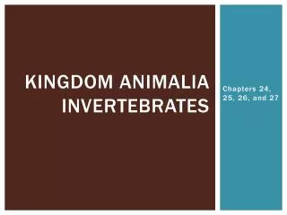
Phylum Cnidaria • Representative Organisms: jellyfish, sea anemones, corals • Evolved tissues to perform specific functions • Swimming, responding to external stimuli, engulfing prey, etc.
Structure • Radial symmetry (oral & aboral surface) • Digestive cavity with one opening (Centrally located mouth • Tentacles to capture & handle food • Cnidocyte – stinging cell containing nematocyst • 3 layers of cells: • Epidermis (external) • Mesoglea (middle gel layer) • Gastrodermis (internal, lining gut)
2 Forms: Polyp & Medusa • Polyp – attached stage with mouth and tentacles oriented upward • Medusa – bell-like upside down polyp specialized for swimming
Feeding & Digestion • Carnivores • Cnidocytes with nematocysts used to capture prey • Food ingested • Digestion in gut • Eliminated quickly
“Poison Darts” – triggered chemically by contacting flesh of an organism Figure 1. A diagram of an undischarged nematocyst in a cell (left). A diagram of the structures found in a nematocyst (right).
Did you know? • The Box Jellyfish is one of the most deadly Jellyfish. In Australia, they kill up to 65 people a year. • The Australian box jelly is responsible for more human fatalities than shark attacks.
Box Jellies • When a Box Jellyfish stings a person, they can die in less than three minutes. • “You have virtually no chance of surviving the venomous sting, unless treated immediately. The pain is so excruciating and overwhelming that you would most likely go into shock and drown before reaching the shore.”
Treatment of Stings • “Vinegar will inactivate undischarged jellyfish stinging cells. The toxin will also help to decrease the symptoms of jellyfish sting. Pouring vinegar on the tentacle before you remove it deactivates the jellyfish stinging cells so more don't fire as you remove it.” • “Urine causes the undischarged stinging cells to fire causing additional stings. “
Behavior • Nerve net transmits impulses • Lack brain / true nerves • Primitive eyes Eyespots of Box Jellyfish (known as a cubazoan)
Moon Jelly Reproduction Juvenile Moon Jellies budding Adult Moon Jelly
Types: Hydrozoans • Feathery colonies of polyps attached to surfaces • Examples: • Siphonophores • Hydra
Examples (cont.): • Portuguese Man-of-War • Blue sail-like float • Transparent tentacles (up to 165 ft long) • Painful sting from toxins in nematocysts
Types: Scyphozoans • “True Jellyfish” • Large medusa (dominant life form) • May reach diameter of 6 ft • Examples: • Moon Jellyfish • Sea Nettle
Types: Anthozoans • Solitary or colonial polyps (lack medusa) • Examples: • Sea anemones • Corals (calcium carbonate skeleton)
http://www.pbs.org/kcet/shapeoflife/episodes/move_explo2.htmlhttp://www.pbs.org/kcet/shapeoflife/episodes/move_explo2.html
B. Diagram 2 Diagram 1 B. A. • The structure labeled A is the ______________________________ of the jellyfish. • The structures labeled B are the ___________________________________________. • These structures contain stinging cells called _____________________________. • In each of these cells, a ____________________ can be triggered to release venom. • The life stage of diagram 1 is a __________________________. • 2 examples of this form of cnidarian are __________________ & ________________. • The life stage of diagram 2 is a __________________________. • 2 examples of this form of cnidarian are __________________ & ________________. • Identify the 3 layers of cells of a cnidarian: external = ______________________________, • middle gel layer = _______________________, & internal = _________________________
Label the life stages above: Adult medusa, Young medusa, Larva, Fertilized egg, Polyp, Polyp transforming
Bilaterally Symmetrical Worms • Bilateralism allows more active pursuit of prey and sophisticated behaviors
Flatworms (Phylum Platyhelminthes) • Simplest bilateral body plan • Flat back & belly • Central nervous system • Simple brain (nerve cells) • Nerve cords • Coordinates movement • 3 cell layers • Epidermis (external) • Mesoderm (middle – muscles, reproductive system) • Internal
Flatworm examples: • Turbellarians – free living carnivores • Flukes – parasites (common in fish, seabirds) • Tapeworms – parasites with long body of repeating units (attached to intestine wall)
Ribbon Worms • More complex than flatworms: • Complete digestive tract (gut with 2 openings) • Circulatory system (blood transports nutrients and oxygen to tissues) • Proboscis – long tube from mouth used to capture prey
Nematodes • Slender cylindrical bodies with pointed ends • Hydrostatic skeleton - Muscle/fluid system • Provides support • Aids in locomotion • Diverse lifestyles: • Parasites • Bottom dwellers
Segmented Worms (Phylum Annelida) • Segmentation: series of similar compartments • Coelom: true body cavity • Closed circulatory system • Blood remains in vessels to transport nutrients, oxygen, and carbon dioxide
Annelid example: Polychaetes • Parapodia – flattened extensions from segments • Gills – absorb oxygen, release carbon dioxide
Lifestyles: • Benthic • Tube worms • Build tubes of mucus, protein, mud, sand • Filter feed (cilia & mucus catch matter) • External surface of sea stars/urchins
Lifestyles (continued): • Hydrothermal Vents – heat & hydrogen sulfide are toxic to most organisms • Lack mouth and gut • Chemosynthetic bacteria in body use sulfide to make food which is used by worms
Plume – captures oxygen, carbon dioxide, and hydrogen sulfide Hemoglobin – binds sulfide and carries thru bloodstream to bacteria Trophosome – feeding body which contains bacteria (285 billion per oz.) Hydrothermal Tubeworm Structure
Annelid example: Leeches • Bloodsuckers attach to fish & invertebrates • Lack parapodia
Agenda – February 26 • Field Trip Materials??? • Invertebrates – Mollusc Notes • Open Notes Quiz – Wednesday • Movie & Questions - VENOM • Test - Friday • Missing Work due by Friday!
Phylum Mollusca • There are more species of molluscs in the ocean than of any other animal group!