Discovering Your Brain: Organization, Function & Dysfunction
460 likes | 563 Vues
Explore the intricate organization, function, and dysfunctions of the human brain, including neural tissue and location descriptions, nervous system divisions, and lateralization of brain function. Learn about brain specialization, plasticity, and investigative brain imaging techniques throughout history to present day. Delve into the fascinating world of brain science!

Discovering Your Brain: Organization, Function & Dysfunction
E N D
Presentation Transcript
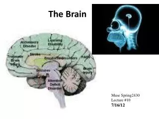
Organization of the Brain • How can we describe the brain? • Neural tissue description(the look) • Location description(the place) • NAT GEO photo gallery
General divisions of the nervous system • Peripheral Nervous System (PNS) • Somatic Nervous System • Autonomic Nervous System • Sympathetic Nervous System • Parasympathetic Nervous System
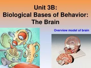
Central Nervous System (CNS) • Spinal Cord • The Brain • Reptilian Brain • Old Mammalian Brain • New Mammalian Brain (Neocortex)
Localization of function of the human brain • Structure and Function, Structure and Function • Subcortical areas • Structure and Function • Structure and Function • Structure and Function
Neocortex • The look • The lobes
A review • NAT GEO web site for review
Let’s Look Into Your Brain! • Today’s topics • Right/Left Brain • Dysfunctions
Lateralization of function of the human brain • Split Brain- although similarly located both cerebral hemispheres generally have similar functions, but. . . • There are some differences or lateralization of functions shown to exist • How did scientist figure this out???
How is this studied? • Electrical stimulation • PET scans • Cerebral vascular accidents (strokes), injury or lesioning • Left/Right Side neglect • Split brain patients • Drugs affecting half of brain • Dichotic listening
Roger Sperry • According to Dr.Sperry, the brain has two hemispheres with different but overlapping functions. The right and left hemispheres of the brain each specialize in distinct types of thinking processes.
Left Hemisphere Specialization • Speech and Language Functions • Wernicke’s Area • Broca’s Area
Right Hemisphere Specialization • Spatial Functions (patterns), visual configurations, color discrimination) • Musical Functions
Hemispheric Dysfunction • Broca’s Aphasia • Wernicke’s Aphasia • Anomic Aphasia • Global Aphasia • Developmental Dyslexia
Plasticity • Chudler/plasticity • The Brain 7 mins
Brain Dysfunction • http://www.ted.com/talks/jill_bolte_taylor_s_powerful_stroke_of_insight.html • Ted talks and strokes
Investigative Assignment • Brain Imaging and web search • Complete prior to slides 52- 70
Windows to the Brain • The Greeks • Franz Gall • Brain Damage – Phineas Gage\ • Walter Hess • Lesion (ablation) • Imaging Techniques
The Greeks Hippocrates Galen 130-200 CE Fluids in the brain ventricles were responsible for sensations, reasoning, judgment and memories • 460-377 BCE • Emotions, thought and mental health arise from the brain
What is Phernology • Phrenology was a faculty psychology. In layman’s terms – reading head features, size, & bumps to determine characteristics, personality, and intellect. • Phrenology was derived from the theories of the idiosyncratic Viennese physician Franz Joseph Gall (1758-1828). • The basic tenets of Gall’s system: • 1. The brain is the organ of the mind. • 2. The mind is composed of multiple distinct, innate faculties. • 3. Because they are distinct, each faculty must have a separate seat or "organ" in the brain. • 4. The size of an organ, other things being equal, is a measure of its power. • 5. The shape of the brain is determined by the development of the various organs. • 6. As the skull takes its shape from the brain, the surface of the skull can be read as an accurate index of psychological aptitudes and tendencies.
Phineas Gage A railroad construction worker in Vermont in 1848. Dynamite blew a tamper rod through his eye and out his skull. Remarkably, he survived, but was never the same. Once considerate and friendly, he was now overbearing, inappropriate, and indecisive. What did we learn from Phineas Gage? We developed a theory about the frontal lobe and we determined that the executive control system in prefrontal cortex was damaged.
Walter Hess • Studied brain function. First to discover function of hypothalamus and “start-stop eating” function • 1955
Lesions • Cutting of brain tissue • Ablation – destroy brain tissue • (Essentially these are the same thing)
Brain Imaging Techniques • The are now many, highly specific methods to see into your brain. • Assignment yesterday helped you examine these. • As a review. . .
More modern approaches • EEG • CAT • MRI and fMRI • PET • MEG • SPECT • DTI
EEG • Transmit electrical activity (brain waves)
CAT or CT scan • Computerized axial tomography- computerized image of x-rays
MRI • Magnetic resonance imaging – magnetic field to develop image of the brain
fMRI • Functional MRI – detect the use of oxygen in the brain
PET • Positron emission tomography- slightly radioactive solution injected to see metabolic activity in imaged part of the brain
MEG • Magnetoencephalography- detect activity too brief to be detected by PET or MRI
SPECT • Single photon emission computerized tomography- tracks cerebral blood flow as an indicator of neural activity in a specific region of the brain
Diffusion Tensor Imaging • An MRI technique that measures the diffusion of water within a cell to yield an image of axons and neural tracts