Download
1 / 75
760 likes | 1.02k Vues
Introduction to Minor Surgical Procedures. Tammy Pifer Than, MS, OD, FAAO UAB School of Optometry tthan@uab.edu. Minor Surgical Procedures in Optometry???. Punctal Occlusion Dilation & Irrigation Cyst incision and evacuation Corneal debridement Corneal foreign body removal

E N D
Introduction to MinorSurgical Procedures Tammy Pifer Than, MS, OD, FAAO UAB School of Optometry tthan@uab.edu
Minor Surgical Procedures in Optometry??? • Punctal Occlusion • Dilation & Irrigation • Cyst incision and evacuation • Corneal debridement • Corneal foreign body removal • Anterior stromal puncture • Chalazion incision and curettage • Papilloma Removal • Correction of Trichiasis • Thermal punctal cautery
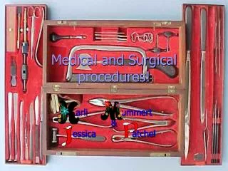
Tools of the Trade • Westcott scissors • iris scissors • tissue forceps • chalazion clamp • curette(s) • Jaeger plate
Instrument Sterilization • Autoclave • a must for intraocular surgery • Ethyl Oxide Gas • alternative to autoclave • Chemical Germicide • destroys most bacteria and viruses • may not eliminate spores • adequate for minor extraocular procedures • Follow manufacturer’s guidelines
Asepsis • Defined: the prevention of contact with microorganisms; freedom from infection • Aseptic techniques • disinfecting surgical area • establishing a sterile field • proper instrument handling • wiping down work areas with germicide or diluted bleach (1:10)
Infection Control • Protocols by CDC and OSHA • Universal Precautions • hand washing • donning gloves • protective eyewear, masks, gowns
Biohazardous Waste and Sharps • Biohazardous Waste • “any material other than sharps that is contaminated with blood, other body fluids, or tissue” • dispose in proper containers according to OSHA • Needles and other sharps go in proper container • NEVER recap a used needle • Blood spills can be disinfected with 1:10 bleach
Laboratory Affiliation • Blood Work (phlebotomy) • Send samples • Send patient • Pathology evaluation • excised lesions
Medicolegal Considerations • Informed Consent • written • Standard of Care • Chart Documentation • before • during • after
Procedure Safety Precautions • Office Protocols • Preoperative vitals • Blood pressure • Pulse • Temperature • Informed consent • Written! • Postoperative instructions • contact numbers
Getting Prepared… • Injection Techniques • Local Anesthesia • Lidocaine 1% or 2% • 1 cc tuberculin syringe (25 G ½”) • Can you sew?
Concretion Removal • “Lithiasis” • removal not always indicated • one drop of topical anesthetic • ± vasoconstrictor • use 25 gauge needle to remove • one drop of topical antibiotic
Sebaceous Cyst Removal • well-demarcated • non-inflammatory • creamy white if superficial • skin-toned if deeper • excise for cosmesis
Sebaceous Cyst Removal • clean skin with alcohol • patient fixates away from cyst • Lidocaine injection • local infiltrative • pull skin taut • score the top of the cyst with a scalpel (cut away from the eye!)
Sebaceous Cyst Removal • use cotton swabs to evacuate lesion • best to cauterize wall of cyst • antibiotic ung qid x 1 week
Sudoriferous Cyst Removal • retention of sweat glands • clear, fluid-filled • remove for cosmetic reasons • direct patient’s gaze away from lesion • use tip of 25 gauge needle to puncture cyst • use cotton swab to collect clear exudate • apply antibiotic ointment in office
Inclusion Cyst Removal • “blister” of the conjunctiva • clear if epithelial • opaque if epi and goblet cells • precipitating factors: • trauma • surgery • foreign body • inflammation
Inclusion Cyst Removal • topical anesthetic • puncture cyst with sterile needle • massage through closed lids • topical antibiotic • massage x 1 week • usually recur
Milium • 1-2 mm epidermal cyst • white - yellow • sites: eyelids, cheeks, forehead • treatment: • incision and expression
“Make it go away!” • 63 YOF • CC: “I want to get rid of this bump” • OHx: Lesion is probably benign • long standing • no change • uniform color • < 6 mm • no bleeding
Squamous Papilloma • aka skin tags or acrochordons • epidermal hyperplasia • skin-colored or hyperpigmented • F > M • one or many • often pedunculated • sites: neck, axilla, eyelids
Papilloma and Verruca Removal • cosmesis • biopsy • visual disturbance
Remember your H-ABCs • H: hair, history • A: asymmetry, avascular • B: borders, bleeding • C: color, change • S: size • If unsure -> send for histopathological analysis • Stay within your comfort zone
Papilloma Treatment Options • leave it alone • chemical cautery • argon laser removal • surgical excision • lesion can be sent for histopathological analysis • requires local anesthesia • pedunculated may be an exception • Stay within your comfort zone!
Papilloma Removal: Informed Consent • Potential Complications • scarring • lid notching • infection • recurrence • Get it in writing!! • Make sure patient is not a keloid former!
Papilloma Removal: Procedure • topical anesthetic OU • ± sterile drape • clean area • local infiltrative injection of lidocaine • use Jaeger plate if near globe • inject ~0.2 cc
Papilloma Removal: Procedure • grasp lesion with tissue forceps • remove at base with scissors or scalpel • place lesion in fixative (if sending to lab) • cauterize • antibiotic ung
Papilloma Removal: Post-Operative • Patient Education • antibiotic ung x 1 week • scab in 1-2 weeks • red area 6-8 weeks • RTC 1 week
Chalazion • benign lesion • sterile lipogranulomatous inflammatory lesion • can cause visual disturbances • measure size • determine if it is anterior or posterior to the tarsal plate
Chalazion: Management • warm compresses • DIGITAL MASSAGE • Many will resolve • oral antibiotics are not indicated unless…
Chalazion: Intralesional Steroid Injection • Hx: how long has it been there? • review complications: • depigmentation • recurrence • infection • ineffective • written, informed consent
Chalazaion: Intralesional Steroid Injection • Procedure • topical anesthetic OU • swab conjunctiva with xylocaine 4% • apply chalazion clamp • inject Kenalog 40 INTO lesion • massage • RTC 2-3 weeks
Incision and Curettage • Patient Preparation • Potential complications • scarring • lid notching • recurrence • loss of cilia • permanent gland obstruction • written, informed consent
Incision and Curettage • Procedure • determine if skin or conjunctival approach • topical anesthetic OU • +/- sterile drape • swab conjunctiva with lidocaine 4% • apply chalazion clamp • inject with lidocaine for local anesthesia
Incision and Curettage • Procedure • make incision with scalpel • skin: horizontal • conjunctival: vertical • scoop out contents with curette • remove capsule wall and cauterize • may inject steroid • control bleeding
Incision and Curettage • Procedure • suture if cutaneous approach • interrupted sutures • usually 3 or 4 • antibiotic ung x 1 week • remove sutures in 3-5 days • RTC 1 week post-op
Corneal Debridement • enhances epithelial healing • removes replicating virus • indications: • recurrent corneal erosions • traumatic corneal abrasions • corneal burns • herpes simples keratitis (epithelial)
Corneal Debridement: Procedure • Instill anesthetic • pull epi towards center of defect • scrub basement membrane
Corneal Debridement: After… • If not HSK… • cycloplegic agent • antibiotic ointment • ± pressure patch • RTC 24 hours; then 3-4 days • IF HSK… • cycloplegic agent • antiviral and antibiotic • NO patch • RTC 1 day
Anterior Stromal Puncture • promotes firm adherences of epithelium • used for recalcitrant RCE • instill anesthetic • debride area? • apply 20-50 punctures into anterior stroma • Beyond defect
ASP: After… • cycloplegic agent • antibiotic ointment/solution • pressure patch or bandage CL • pain management • RTC 1 day • LONG TERM USE OF HYPERTONICS!
Suture Removal: Cutaneous • Interrupted Suture • lift sutures with forceps • cut suture just above skin • pull knotted end towards wound • do not drag exposed suture through wound
Suture Removal: Cutaneous • Running Suture • Cut every other strand at skin surface • grap middle portion and pull • remove knots as interrupted sutures
LASER • Light • Amplification by • Stimulated • Emission of • Radiation
Properties of Laser Light • Monochromaticity • UV 40 - 379 nm • Visible 380 - 760 nm • IR 760 - 4x10⁵ nm • High Power Density • energy = number of photons • power = energy / sec • power density = energy / sec / area
Laser-Tissue Interactions • the effect on physiologic tissue when exposed to laser light • can alter by changing laser variables • wavelength • exposure time • spot size
Laser-Tissue Interactions • Photocoagulation • Photovaporization • Photodisruption • Photoablation • Photoasepsis • Photodynamic • Photostimulation