Download
1 / 51
510 likes | 900 Vues
DISORDERS OF THE IMMUNE SYSTEM: ALLERGIES. Your Immune System. The organs involved with the immune system are the lymphoid organs, which affect growth, development, and the release of lymphocytes.

E N D
Your Immune System The organs involved with the immune system are the lymphoid organs, which affect growth, development, and the release of lymphocytes. The blood vessels and lymphatic vessels are important parts of the lymphoid organs, because they carry the lymphocytes to and from different areas in the body Each lymphoid organ plays a role in the production and activation of lymphocytes. Lymphoid organs include: adenoids, tonsils, appendix, lymph nodes, spleen, Peyer’s patches (lymph tissue in your small intestine), thymus
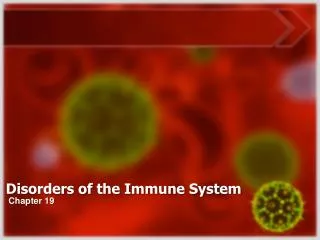
Disorders of the immune system • When the immune system does not function properly, it leaves the body susceptible to an array of diseases • Allergies and hypersensitivity to certain substances are considered immune system disorders • Examples of immune disorders include: • cancer of the immune system (leukemia) • autoimmune diseases, such as juvenile diabetes, rheumatoid arthritis, and anemia • immune complex diseases, such as viral hepatitis and malaria • immunodeficiency diseases, such as acquired immunodeficiency syndrome (AIDS)
Autoimmune diseases • autoimmune rx’s can be directed against • the brain multiple sclerosis • the gut in Crohn's disease • other autoimmune diseases such as systemic lupus erythematosus (lupus) • damage to certain tissues by the immune system may be permanent, as with destruction of insulin-producing cells of the pancreas in Type 1 diabetes mellitus
Hypersensitivity • Abnormal sensitivity to microorganisms, products or certain foreign substances • Sensitivity beyond what is considered normal
Nature of Hypersensitivity • Chemical nature of the allergen (pollen, food, latex, metals, synthesized chemicals, penicillin) • Sensitizing route-ingestion, injection, skin contact • Individual traits-anatomical and physiological traits • Once sensitized, another exposure to the Ag triggers an immune response that damages host tissue –sensitization can takes years I.e. a lot of people come to AZ with NO allergies, after 5-7 years they develop allergies
Allergen • Any substance which gives rise to allergy (antigen)
HYPERSENSITIVITY (allergy) • Excessive (harmful) immune response • Individuals previously exposed (sensitized) to an antigen (allergen) • Four main types (I-IV)
TYPES OF HYPERSENSITIVITY • Type I • Anaphylatic reaction (less than 30 min) • Type II • Cytotoxic reaction (5 to 12 h)
TYPES OF HYPERSENSITIVITY (cont.) • Type III • Immune-complex reaction (3 to 8 h) • Type IV • Cell-mediated reactions (24 to 48 h)
TYPE I (ANAPHYLATIC REACTION) • Ana (against), phylaxis (protection) • Hay fever • Asthma • Hives
Type I (mechanism) • Antigenic stimulus (Ag’s + Ig’s combine) • IgE production is triggered • IgE’s might trigger a systemic response, producing sometimes fatal shock and breathing difficulties • or a localized rx, such as hay fever, asthma or hives • IgE bind to mast cells and basophils • Mast cells are prevalent in the connective tissue of skin, the respiratory tract, and surrounding blood vessels • Basophils circulate in the blood
Type I (mechanism) (cont.) • Mast cells and basophils degranulate after successive antigenic stimuli • Histamine --affects blood vessels-causing edema, erythema, increased mucus • secretion and smooth muscle contraction, resulting in breathing difficulties • Leukotrienes –cause contraction—like the spasms of asthmatic attacks • Prostaglandins tend to cause increased secretion of mucus
Type I (local reactions) • Hay fever (histamine-mediated response to inhaled antigen) • Asthma • Inhalation • Hives (urticaria) • Ingestion of food or drug—often difficult to distinguish btw food hypersensitivity & food intolerance
Urticaria wheals on the thigh Swelling of lips and eye lids
Can hives occur anywhere else?Around 1 in 3 people with hives can also have swelling of the tongue and throat. This is called angioedema, and is caused by similar swelling deeper in the tissues. Occasionally the swellings will occur inside the stomach and cause tummy pain or cramps. Underneath the lining of the skin, gut, lungs, nose and eyes are mast cells. These are designed to kill worms and parasites. Mast cells are like "land-mines", and contain "bags" filled with irritant chemicals including histamine. When these are released in small amounts, they cause local itch and irritation. In larger amounts, they will cause fluid to leak out of blood vessels, resulting in swelling of the skin.
Allergic reactions to food, pain killers (such as aspirin or arthritis tablets like naproxen, diclofenac) or antibiotics can also trigger hives. Sometimes insect stings, food additives or preservatives can trigger hives
TYPE I (systemic reactions) • Anaphylactic shock • Injected antigens (bee sting) • weak pulse,low blood pressure, low temperature-- it may be fatal • Breathing difficulties
PREVENTION OF ANAPHYLACTIC REACTIONS • Avoid contact with the allergen • Skin tests (wheals) • Determine cause • Desensitization (injection of small doses of the Ag—to induce IgG AB’s production, to serve as blocking AB’s that intercept & neutralize Ag’s before they can react with cell bound IgE’s)
TYPE II (cytotoxic reactions) • Cell (RBC or tissue cells) bearing or coated with antigens • Antibody-antigen reaction activates complement • Complement & MQ destroys antigen bearing cells
TYPE II (cytotoxic reactions) • Transfusion reactions • ABO blood group system • Carbohydrate antigens A and, or B on RBC membrane • O group lacks antigen Anti B plasma AB’s Anti A plasma AB’s Type O Type A Type B Can receive type A & O only Can receive type B and O only
Innocuous antigens can cause type II hypersensitivity reactions in susceptible individuals by binding to the surfaces of circulating blood cells AB-mediated destruction of RBC (hemolytic anemia) or platelets (thrombocytopenia) is an uncommon side-effect associated with the intake of certain drugs such as the antibiotic penicillin, the anti-cardiac arrhythmia drug quinidine, or the antihypertensive agent methyldopa. These are examples of type II hypersensitivity reactions in which the drug binds to the cell surface and serves as a target for anti-drug IgG antibodies that cause destruction of the cell. The cell-bound antibody triggers clearance of the cell from the circulation, predominantly by tissue macrophages in the spleen, which bear Fc receptors
TYPE II (cytotoxic reactions) (cont.) • Blood type hereditary pattern • Roughly 85% of the human population has an Ag named Rh factor and they are called Rh +
TYPE II (cytotoxic reactions) (cont.) • Antibodies against A and B blood antigens are naturally present without previous stimulation, they are type IgM
TYPE II (cytotoxic reactions) (cont.) • Rh blood group • Antigen present in 85% of the population (Rh+) • Antibodies against Rh blood group appear only after exposure of Rh- individuals to Rh+ blood (pregnancy) • Antibodies against Rh blood group are type IgG
TYPE II (cytotoxic reactions) (cont.) • Hemolytic disease of the newborn • (erythroblastosis fetalis)-destruction of RBC’s • Rh- mother pregnant with an Rh+ baby • Transfusion of an Rh- mother with Rh+ blood
TYPE II (cytotoxic reactions) (cont.) • Mother develops anti Rh+ antibodies • Later pregnancies with Rh+ babies, antibodies (IgG) cross the placenta and destroy baby’s RBC
PREVENTION OF ERYTHROBLASTOSIS FETALIS • Passive immunization of the mother with anti-Rh+ antibodies immediately after childbirth • Transfusion of fresh blood to the baby
Drug Induced Cytotoxic Reactions Thrombocytopenic purpura occurs when blood platelets, which are essential for clotting, are coated w/ drug molecules that function as haptens If Ab’s develop against these haptens cause destruction of the platelets In hemolytic anemia, the body may form AB’s against its own blood cells Immune-caused destruction of WBC’s is called agranulocytosis
TYPE III (IC reaction) • This is caused when soluble antigen-antibody (IgG or IgM) complexes, which are normally removed by macrophages in the spleen and liver, form in large amounts and overwhelm the body • These small complexes lodge in the capillaries, pass between the endothelial cells of blood vessels - especially those in the skin, joints, and kidneys - and become trapped on the surrounding basement membrane beneath these cells • IC circulate in the blood • Pass between blood vessel cells (endothelial) • The antigen/antibody complexes then activate the classical complement pathway and cause inflammation
TYPE III (Immune complex rx’s) • Antibody-soluble antigens (immune complexes) in serum escape phagocytosis b/c they are too small • These circulating AB-AG complexes get deposited in organs and can cause inflammation and damage • Glomerulonephritis is an immune complex condition that causes inflammatory damage to kidney glomeruli
TYPE III (IC reaction) (cont.) • Glomerulonephritis • Is caused by the response of the immune system to the M protein of Streptococci pyogenes (Group A Streptococci—GAS) • May lead to a fatal kidney failure
What is glomerulonephritis? -Glomerulonephritis is the term used to describe a group of diseases that damage the part of the kidney that filters blood -other terms used are nephritis and nephrotic syndrome
Acute glomerulonephritis • develops suddenly • may get it after an infection in your throat or on your skin (Streptococcus-- M protein) • The early signs of the acute disease are • puffiness of your face in the morning • blood in your urine (or brown urine) • urinating less than usual
Rheumatoid • Arthritisis an • Auto Immune rx • IC are • deposited • in the joints • -it is a systemic disease • In RA, the inflammatory • process continues on, • creating excessive • inflammation that • can cause damage
TYPE IV (Cell mediated reactions) • Type I-III hypersensitivities involved IgE, IgG or IgM • Type IV rx’s involve cell-mediated rx’s caused by T cells/or MQ • Delayed reactions (24-48 h) • During the delayed rx-- MQ and lymphocytes migrate & accumulate near the foreign AG • Foreign Ag is phagocytized by MQ—presented to T cell surface receptors • T cells involved in this type of rx are TD but may include Tc ells • A principal factor is the release of lymphokines by T cells rx w/ the target Ag • TD and TC cells • Two examples (PPD test and poisen ivy)
Tuberculin Hypersensitivity. • observed when soluble AG from organisms such as Mycobacteria bovis (PPD test)were administered subcutaneously • fever, general unwellness, plus an area of red, hard swelling • This type of rx is induced by a series of cellular migrations and activations • T-cell migration from capillaries • Disruption of collagen in dermis • Macrophage infiltration • Granulomatous appearance, no edema, self-limiting
What is a granuloma? • When macrophages fail to destroy the mycobacteria, the human immune systems next line of defense is to form granulomas around the infected macrophages • Layers of two different types of T cells surround the infected macrophage, sealing it inside a barrier from which it cannot escape • This barrier is known as a granuloma • Since the infecting mycobacteria is "contained" inside the granuloma, this form of mycobacterial disease is known as the contained form. Contained mycobacterial disease is typified by low numbers of infecting mycobacteria and high levels of inflammation
PPD skin test • T cells are responsible for the formation of these granulomas • PPD stands for Purified Protein Derivative. The subject mycobacteria are killed, using heat or ultrasound • PPD for every mycobacterium is different. • PPD for M. tuberculosis is called "tuberculin", that for Mleprae is called "lepromin" or "leprosin • site of injection is then checked one or two days later. • skin around the injection site is inflamed, or has formed a granuloma, then this shows that the patient is currently infected with the mycobacterium • Because the reaction only shows up after a day or two, this type of reaction is called Delayed Type Hypersensitivity (DTH)
TYPE IV (Cell mediated reactions) • Re-exposure to antigen • Memory T cells activate • Release of cytokines when in contact with the AG • Allergic contact dermatitis are usually caused by haptens that combine w/ proteins in the skin • Typical foreign AG are poisen ivy, cosmetics, latex and metals such as nickel in jewlery • The patch test, in which samples of suspected material are taped to the skin, may determine the offending environmental factor