Immunoglobulins
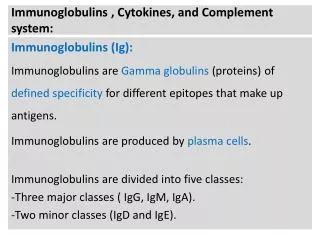
Immunoglobulins. Structure. Definitions. Immunoglobulin is a generic term that refers to a diverse group of molecules found in the blood and tissue fluids They are soluble globulin molecules and they generally migrate in an electrophoretic field at or near the gamma globulin fraction


Immunoglobulins
E N D
Presentation Transcript
Immunoglobulins Structure
Definitions • Immunoglobulin is a generic term that refers to a diverse group of molecules found in the blood and tissue fluids • They are soluble globulin molecules and they generally migrate in an electrophoretic field at or near the gamma globulin fraction • An antibody is an immunoglobulin molecule capable of binding specifically with a known substance (antigen) • Immunoglobulins are synthesized by B lymphocytes and terminally differentiated B lymphocytes or plasma cells
Introduction • The first to show that antibodies reside in the gamma globulin fraction were Kabat and Tiselius in 1939 when they performed serum electrophoresis and they called them immunoglobulins • From the broad electrophoretic peak, it is clear that a heterogeneous collection of immunoglobulin molecules with slightly different charges is present • This heterogeneity was one of the early obstacles in attempts to determine the structure of antibodies, since analytical chemistry requires homogeneous crystalizable compounds as starting proteins
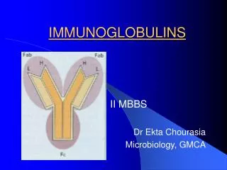
Certain diseases (multiple myeloma and light chain disease) provided the solution • Recently the hybridoma technology has made available antibodies in pure form • Porter in 1959 treated antibody with papain and produced three fragments of equal size • Two of the fragments were found to retain the antibody’s ability to bind antigen specifically but could no longer precipitate the antigen from solution • These two fragments were referred to as Fab • The third fragment could be crystallized out of solution and called FC
ENZYME DIGESTION OF IMMUNOGLOBULINS Fragment “antigen-binding” Papain Fragment crystallisable Pepsin
At about the same time, Edelman discovered that when the immunoglobulin molecule was extensively reduced of by mercaptoethanol, it fell apart into four chains, two identical heavy chains (53000 Daltons) and two identical light chains (22000 Daltons) • Noble prize was awarded for Porter and Edelman for revealing antibody structure
Antibody structure LIGHT HEAVY H+L Antibody + dimer
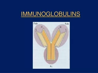
Structure • An immunoglobulin is composed of four polypeptide (glycoprotein) chains; two light and two heavy chains linked by disulfide bonds • Five different types of heavy chains; α, , µ, δ, and ε. • Two types of light chains; kappa (k) and lambda(λ) with a ratio of 3:2 • An individual B cell or immunoglobulin monomer expresses either k or λ but not both • Within an immunoglobulin molecule, the two heavy chains and light chains are identical
Some immunoglobulin molecules are basic monomers and others are composed of multiple copies (dimers or pentamers) of identical immunoglobulin monomers • Both heavy and light chains can be divided into regions or domains, homologous portions of an immunoglobulin chain, each composed of approximately 110 amino acids and contains an intradomain disulfide bridge
(b) The μ and Є heavy chains contain an additional domain that replaces the hinge region • Heavy and light chains are • folded into domains, each containing • about 110 a.a. & an intrachain disulfide bond • that forms a loop of 60 amino acids.
Light chains contain two regions, a variable (VL) and a constant ( CL) domain • Heavy chains contain a single variable (VH) and multiple constant domains(CH1, CH2, CH3, CH4) • Variable regions in both heavy and light chains are so named because of the extensive variation in the amino acid sequence found in immunoglobulin molecules made by different cells • The amino acid sequence determines the conformational structure of VH and VL • The combination of a light variable and a heavy variable region forms a pocket that constitute the antigen-binding region of the immunoglobulin molecule
Hinge Region • Made up predominantly of cysteine and proline residues • It permits flexibility between the two Fab arms of the Y shaped molecule • It allows Fab to open and close to accommodate binding to two epitopes separated by a fixed distance
Variable Domains • Three areas of hypervariability occur between less variable stretches called Framework regions • Because they bind specifically to epitopes, the hypervariable regions are termed complementarity -determining regions (CDRs) of the L and H chains; CDR1, CDR2, and CDR3
V CDR1 CDR2 CDR3 V D J J CDR1 CDR2 CDR3
Immunoglobulin Fragments • Fab: produced by papain cleavage (2 Fab) • FC: produced by papain cleavage • Fd: it is the heavy chain portion of an Fab fragment cleaved by papain • F(ab)2: it is the dimeric molecule produced by pepsin cleavage which fragments the FC • Fd─: it is the heavy chain portion of an Fab fragment cleaved by pepsin
Immunoglobulin Classes • Five different classes or isotypes depending on heavy chain antigenicity • Monomers, dimers or pentamers • Monomers are divalent having two identical antigen- binding sites
General structures of the five major classes of antibody
IgM • First to be produced in an immune response. M for macroglobulin, M. Wt. about 106 Daltons, sedimentation coefficient 19S, Has an extra CH domain • Cell surface bound monomer or secreted pentamer. Five basic units (pentamer) joined by a J chain (150.000 Daltons) synthesized by B cells or plasma cells • Five antigen-binding sites instead of the expected valence of 10 due to conformational constraints imposed by polymerization • 6-8% of serum immunoglobulins
IgG • Monomer, M. Wt 150.000, sedimentation coefficient 7S. Least anodic of all serum proteins • Four subclasses; IgG1, IgG2, IgG3, and IgG4 • Predominant immunoglobulin in blood, lymph, CSF, and peritoneal fluid • 72-80% of serum immunoglobulins
IgA • The major immunoglobulin in external secretions such as saliva, mucous, sweat, gastric fluid, and tears • Monomer (serum) or dimer (secretions), M.Wt 165000, sedimentation coefficient 7S • Two subclasses; IgA1 andIgA2 • 13-19% of serum immunoglobulins • Serum IgA is predominantly monomeric and monomeric IgA1 accounts for about 90% of serum IgA
Secretory Ig A • Secretory Ig A has a J chain and a secretory piece (70.000 Daltons) which is synthesized by epithelial cells to facilitate passage of secretory IgA into mucous secretions and to protect it from cleavage (secretory IgA is more resistant than serum IgA) • Secretory IgA: - IgA1 90% in secretions above the diaphragm - IgA2 10% in lower GI
IgD • Monomer, M. Wt. 180.000, sedimentation coefficient 7S • A major surface component of many B cells but present in very low concentrations (< 1% of serum immunoglobulins) in serum where it has no function. • It is not secreted by plasma cells and it is uniquely susceptible to proteolytic cleavage
IgE • Monomer, M. Wt 200.000, sedimentation coefficient 8S • It has an extra CH domain • Less than 0.001% of serum immunoglobulins • Binds with high affinity to mast cells and basophils (Homocytophilic)
Allotypes • Allelic forms of the same protein as a result of the presence of different forms of the same gene at a given locus • Allotypic differences at known loci usually involve changes in only one or two amino acids in the constant region of a chain • Important genetic markers inherited as dominant traits; Gm on chain, Km (previously Inv) on kappa chains, and Am on α chain • The genes encoding the markers are expressed codominantly, so that an individual may be homozygous or heterozygous for a given marker
Ideotypes • The antigenicity of the variable region of Fab • May or may not block binding of the antibody to the antigen depending whether produced against CDRs or Framework sequence. An ideotype represents an “internal immage” of the epitope • Public or cross reacting epitopes are ideotypes on different antibodies produced against the same epitope. Private epitopes react with only a particular antibody molecule • Regulation of an Immune response (Jerne’s Theory)