Download
1 / 45
450 likes | 479 Vues
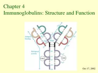
Explore the intricate 3D structure of immunoglobulins, including antibody flexibility, myeloma proteins, and antigen-binding sites. Learn about the common fold, variability, and motion within antibodies.

E N D
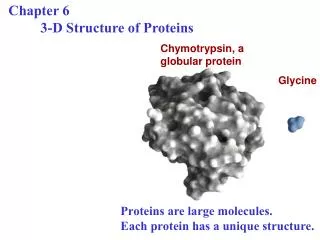
3 D Structure of Immunoglobulins Hugh B. Fackrell Filename:Kuby05b7.ppt
Assigned Reading • Content Outline • Performance Objectives • Key terms • Key Concepts • Short Answer Questions
ASSIGNED READING • Chapter pp
Antibody structure • Special antibodies to study structure • Myeloma proteins • Bence Jones Proteins • Monoclonal Antibodies • 3D structure of antibodies • Binding site • Variability in structure
Mulptiple Myeloma • Disease • Plasmocytoma • Cancer of antibody producing cell • Symptoms • recurrent infections, anaemia, fractures, pain, bone lesions • Detection • Electrophoresis, X-rays, blood smear
Bence Jones Protein • Homogenous light chains • In urine of myeloma patients • Easily purified • Insoluble at 60 -70 oC • Soluble at 80 oC
Monoclonal Antibodies • Add information here
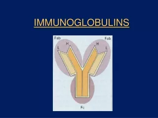
Problems with flexibility • Difficult to determine entire structure • Hinge flexibility • Fragment • Study Fragment • Assemble composite
Bence Jones Light chains • Simplest fragments to isolate • Easily crystallized • Mimics Fab
Immunoglobulin fold • Signature structure common to all immunoglobulins • 2 beta sheets • held by disulfide bond • Variations in detailed structure set specificity
Hypervariable Regions • Discovery • KW plots • Light chains • Heavy chains • CDR vs HVR
Kabat & Wu Plots • Why • demonstrate details of variability of protein sequence of similar proteins • Results • Variability = 1.0 >> no variation • Variability = 400 >> 20 different AA Number different amino acids Variability = Frequency of most common AA
Performance Objectives Key terms, concepts short answers