Download
1 / 1
30 likes | 296 Vues
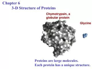
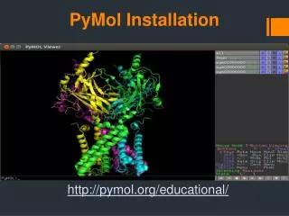
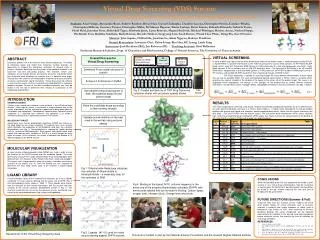
16 blades 2 processors/blade 2 cores/processor = 64 total parallel cores. HP Front end 2.4GHz + 2.4GHz. 128 GB of memory total. 2.4 GHz 2.4 GHz. 2.4 GHz 2.4 GHz. 2.4 GHz 2.4 GHz. 2.4 GHz 2.4 GHz. 2.4 GHz 2.4 GHz. 2.4 GHz 2.4 GHz. 2.4 GHz 2.4 GHz. 2.4 GHz 2.4 GHz.

E N D
16 blades 2 processors/blade 2 cores/processor = 64 total parallel cores HP Front end 2.4GHz + 2.4GHz 128 GB of memory total 2.4 GHz 2.4 GHz 2.4 GHz 2.4 GHz 2.4 GHz 2.4 GHz 2.4 GHz 2.4 GHz 2.4 GHz 2.4 GHz 2.4 GHz 2.4 GHz 2.4 GHz 2.4 GHz 2.4 GHz 2.4 GHz 2.4 GHz 2.4 GHz 2.4 GHz 2.4 GHz 2.4 GHz 2.4 GHz 2.4 GHz 2.4 GHz 2.4 GHz 2.4 GHz 2.4 GHz 2.4 GHz 2.4 GHz 2.4 GHz 2.4 GHz 2.4 GHz 2.4 GHz 2.4 GHz 2.4 GHz 2.4 GHz 2.4 GHz 2.4 GHz 2.4 GHz 2.4 GHz 2.4 GHz 2.4 GHz 2.4 GHz 2.4 GHz 2.4 GHz 2.4 GHz 2.4 GHz 2.4 GHz 2.4 GHz 2.4 GHz 2.4 GHz 2.4 GHz 2.4 GHz 2.4 GHz 2.4 GHz 2.4 GHz 2.4 GHz 2.4 GHz 2.4 GHz 2.4 GHz 2.4 GHz 2.4 GHz 2.4 GHz 2.4 GHz 1 2 3 Fig 2. Ligands (#1-15) used for virtual drug screening against DHFR enzyme 4 5 6 7 8 9 10 11 12 13 14 15 Virtual Drug Screening (VDS) Stream:Can new drugs be identified from virtual libraries of drug-like molecules? Students: Azael Arizpe, Alessandra Rossi, Andrew Ramirez, Bryan Han, Crystal Chaloupka, Claudine Lucena, Christopher Nevins, Candace Whaley, Christopher Millican, Courtney Tanwar, Christopher Miller, Da’Marcus Baymon, Damir Ljuboja, Devin Greene, Eduardo Elizondo, Gabriela Torres, Heidi Held, Jonathan Pena, Abdul-Jelil Tagoe, Kimberly Jones, Liana Renteria, Megan Hardwick, Michael Wissinger, Marissa Acuna, Nathan Hoppe, Phi-Khanh Tran, Radhika Sakalkale, Rathi Kannan, Ricardo Saldivar, Sungryong Lim, Sarah Barnes, Thanh Thao Pham, Yiling Wu, Zoe O’Connor Mentors: John Aquino, Clifford Ho, Jonathan Le, Adam Nguyen, Kathryn Pendleton Alumni Researchers: Lawrence Choi, Dylan Estep, Kim Hsu, KC Leung, Linda Yang Instructors: Josh Beckham (RE), Jon Robertus (PI) Teaching Assistant: Brad Wallentine Freshman Research Initiative, Dept. of Chemistry and Biochemistry, College of Natural Sciences, The University of Texas at Austin VIRTUAL SCREENING In order to dock each ligand into the three dimensional model of the protein target, a software program called GOLD4 is implemented in a parallel architecture on the TI3D Drug Discovery Cluster, which contains 16 HP Proliant BL35P blade servers, each with 2 dual core AMD Opteron 2.4 GHz processors for a total of 64 processors (see Fig 3). Each blade contains 8 GB of memory and a 6 GB ATA hard disk drive. The front-end of the cluster is an HP xw9300 Workstation, equipped with a dual core AMD Opteron 2.4 GHz processor, an NVIDIA Quadro FX4500 graphics card, 4 GB memory, and two 500 GB SATA hard drives that is backed up through a RAID5 network. The GOLD application is capable of scanning through the many different conformations of each ligand that are due to rotatable bonds. For each corresponding docking conformation a Fitness Score is assigned which quantifies the relative strength of the binding of the ligand to the active site of the protein target. This score is an aggregate which incorporates the change in free energies of the binding due to several different chemical interactions: Van der Waals forces, hydrogen bonds, hydrophobic interactions and strain penalties (see Eqn. 1 below). ABSTRACT Enzymatic proteins are at the heart of many disease processes. The ability to effectively target and inhibit their molecular function provides an opportunity to mitigate the deleterious outcomes of disease states in humans. However, identifying new drug leads using traditional methods is an expensive and time consuming process. This research stream uses computers to sift through libraries of chemical structures and predict which ones may bind most effectively to a protein that is a potential drug target. Through this work students are introduced to fundamental features of protein structures and of protein-ligand interactions. Virtual drug screening software is used and the results are then visualized and interpreted with a molecular graphics program. Several of the best candidate molecules can then be tested in the wet lab to determine their efficacy in comparison to the computational predictions. Overall Process for Virtual Drug Screening Download X-ray crystal structure of protein Analyze 3-D Structure in PyMol Fig 3. Parallel architecture of TI3D Drug Discovery computer cluster with processor speeds shown TI3D Cluster Use parallel computing program to ‘dock’ the potential drugs into the protein target Eqn. 1 INTRODUCTION EXAMPLE DISEASE Cancer is the second most common cause of death in the US behind heart disease1. Each type of cancer is essentially a unique disease due to the variety of pathways that are activated or suppressed to bring about aberrant cell growth and functionality. Consequently targeting each individual cancer separately is a daunting task. However, one approach is to inhibit a molecular pathway that is particularly common in all cancers. MOLECULAR TARGET Along these lines, human dihydrofolate reductase (DHFR) was chosen as the drug target for our virtual screening. This protein acts as an enzyme which facilitates the production of tetrahydrofolic acid from the substrate dihydrofolate (see Fig 1). Tetrahydrofolate is required by rapidly dividing cells to synthesize DNA precursors. Since cancer cells divide much more rapidly than ordinary cells, inhibition of DHFR is a useful therapy for many types of cancer. In this research, we sought to compare the binding of 15 different ligands to the DHFR enzyme. RESULTS The initial computational screening using GOLD software found the best binding conformation out of 10 different ones for each ligand listed below and gave it a Fitness Score (Table 1). A higher score corresponds to stronger binding between the enzyme and ligand and, therefore, greater predicted drug activity. Notice that the natural substrates (the folic acids – FOL and FOLH3) have the highest scores. The anti-cancer drug methotrexate (MTX) scores relatively high, while the anti-bacterial drug trimethoprim (TRR) scores low. Figure 4 shows the representation of the binding of MTX and DHFR as shown in PyMol with orange lines depicting bonds. Rank the candidate drugs according to their binding strength Validate actual inhibition of the best ones in the wet lab using enzyme assays MOLECULAR VISUALIZATION An open source software program called PyMol2 was used in order to view the structure of the DHFR enzyme and the candidate ligands. The three dimensional structure files (.pdb), determined by X-ray crystallography, were obtained from the literature at the Protein Data Bank (www.rcsb.org/pdb)3. The images were manipulated to study the primary, secondary and tertiary structure of the protein (Fig. 4). Then the binding site was analyzed to determine the most likely amino acids to be involved in protein-ligand interactions. LIGAND LIBRARY A small electronic library of the 3-dimensional structures for a set of ligands numbered 1-15 was used for docking into the active site of DHFR (Fig. 2 with corresponding names shown in Table 1). These ligands were chosen from the literature for their known interactions with the enzyme and their similarity to the natural substrate, dihydrofolate (shown in Fig. 1). The structural similarities can be see by the presence of two- membered rings similar to the two membered pterin ring system in dihydrofolate. Fig 1. Dihydrofolate Reductase catalyzes the reduction of dihydrofolate to tetrahydrofolate – a necessary step for the synthesis of DNA • CONCLUSIONS • While not providing proof that any compound will inhibit a given enzyme in vivo, virtual drug screening does help the researcher to narrow down the field of best possible ligands that could then be tested in the lab, thereby reducing the time and cost of bringing new drugs to market. • FUTURE DIRECTIONS (Summer & Fall) • Using the skills from this research stream, students will select other protein targets for virtual screening, such as enzymes involved in bacterial and fungal infections or those used in bioterrorism (like anthrax or ricin). After screening for the best predicted binding, the actual compounds will be obtained commercially for validation in the wet lab using light absorbance based enzymatic assays that quantify the level of inhibition for each drug candidate. • REFERENCES • Center for Disease Control, 1995 • The PyMOL Molecular Graphics System. (2008) DeLano Scientific LLC, Palo Alto, CA, USA • H.M. Berman, J. Westbrook, Z. Feng, G. Gilliland, T.N. Bhat, H. Weissig, I.N. Shindyalov, P.E. Bourne, The Protein Data Bank, Nucleic Acids Research2000, 28, 235-242. • Jones, G.; Willett, P.; Glen, R. C., Molecular recognition of receptor sites using a genetic algorithm with a description of desolvation. J Mol Biol1995, 245, (1), 43-53. Fig 4. Binding of the ligand, MTX, (colored magenta) in the active site of the enzyme dihydrofolate reductase (DHFR) with amino acids labeled that are involved in binding. Carbon (gray), oxygen (red), nitrogen (blue). Orange lines are bonds. Researchers of the Virtual Drug Screening class This work is funded in part by the National Science Foundation and the Howard Hughes Medical Institute