Download
1 / 16
160 likes | 195 Vues
Learn about the third level of protein organization, protein folding, motifs, domains, and quaternary structure. Discover how proteins fold, the role of chaperonins, disulfide bonds, and protein denaturation.

E N D
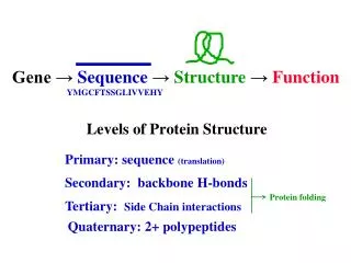
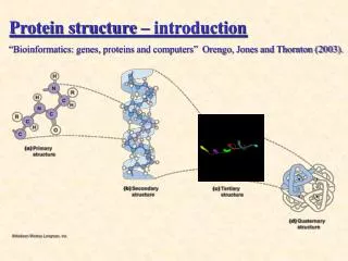
Protein 3-D structure: 3o and 4o structure and protein folding.
3o Structure • third level of protein organization • folding of polypeptide chain causes 2o structures to interact • formation of motifs and domains
Proteins with similar 1o structure also have similar 3o structure tuna 1 GDVAKGKKTFVQKCAQCHTVENGGKHKVGPNLWGLFGRKTGQAEGYSYTDANKSKGIVWNyeast 1 GSAKKGATLFKTRCLQCHTVEKGGPHKVGPNLHGIFGRHSGQAEGYSYTDANIKKNVWDErice 1 GNPKAGEKIFKTKCAQCHTVDKGAGHKQGPNLNGLFGRQSGTTPGYSYSTANKMAVIWEEtuna 61 ETLMEYLENPKKYIPGTKMIFAGIKKKGERQDLVAYLKSATSyeast 61 NNMSEYLTNPKKYIPGTKMAFGGLKKEKDRNDLITYLKKACErice 61 NTLYDYLLNPKKYIPGTKMVFPGLKKPQERADLISYLKEATS
Common Motifs • MotifComposition • Helix-loop-helix all alpha-helix • Coiled-coil • Helix bundle • Beta meander all beta sheet • Greek key • Beta-alpha-beta mixed alpha/beta
Motifs Combine to form Domains • Domains are independent folding units in a 3o structure of a protein • Individual domains have specific function Parallel twisted sheet • Hydrophobic interactions are the major driving force in folding domains Alpha/beta barrel
C O O - C O O - C O O - C O O - + NAD+ O C H O C H + NADH O C H O C H H C C H 2 2 3 C H C H 3 C O O - C O O - Protein family members share common domain structures lactate dehydrogenase malate dehydrogenase + NAD+ + NADH
4o Structure • Quaternary structure describes the organization of subunits in a protein with multiple subunits (oligomeric protein) • Can have homo-multimers or hetero-multimers a2b2 a2bg
4o Structure • Determine molecular weight of native protein by gel permeation chromatography • Determine molecular weight of individual subunits by SDS-PAGE • Can use the information to determine subunit composition If……. Native protein – 160,000 daltons and a-Subunit – 50,000 daltons b-Subunit – 30,000 daltons Then…… Protein can have a2b2 structure
4o Structure • Subunits held together by non-covalent interactions • Oligomeric protein is more stable than disassociated subunits • Active site often made up of AA residues from different subunits • 4o and 3o structure is often affected by ligand (substrate or inhibitor) binding. Important in enzyme regulation
Tm Protein denaturation • Denaturation – disruption of native conformation • Heat commonly used to denature proteins • Tm = temperature where 50% folded/50% unfolded. • Typical Tm = 40-60oC • Tm for thermophiles >100oC (Taq DNA polymerase) • Chemical denaturants Chaotrophic agents = Urea, KCN detergents = SDS
Protein Folding • Ribonuclease A (RNase A) will refold to native structure spontaneously (1 minute) • >1050 possible conformations • If 10-13 sec per conformation would take 1030 years to sample enough to determine structure • How do proteins fold so quickly?
Factors driving protein folding DG = DH - TDS • Conformational entropy A+B C decreases entropy (unfavorable) • Non-covalent interactions give favorable enthalpy value • Hydrophobic effect increases entropy by freeing water (favorable) - +
Protein Folding • Structures of globular proteins are not static • Proteins “breathing” between different conformations • Proteins fold towards lowest energy conformation • Multiple paths to lowest energy form • All folding paths funnel towards lowest energy form • Local low energy minimum can slow progress towards lowest energy form
Pathway of Protein Folding 1) Nucleation of folding - Rapid and reversible formation of local 2o structures form 2) Formation of domains (Molten Globular intermediates) through aggregation of local 2o structures 3) Domain conformations adjust to form native protein
Chaperonins • Protein complexes that promote protein folding • Chaperonins don’t determine native structure • Prevent misfolding and aggregation of protein • Sequesters unfolded protein from other proteins • Require ATP for protein binding, after ATP hydrolysis native protein released • Thought to bind unfolded regions of protein
Disulfides Bonds • Stabilize native structure • Formed after native conformation achieved • Abundant in secreted proteins but not in intracellular proteins • Protein disulfide isomerase catalyzes reduction of incorrect disulfide linkages