Levels of Protein Structure
260 likes | 515 Vues
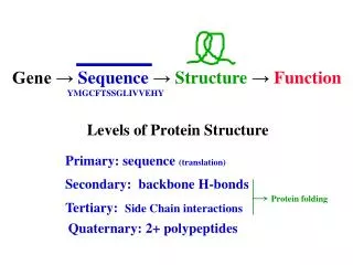
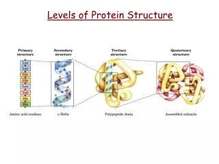
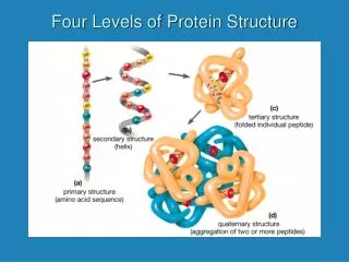
Levels of Protein Structure. Gene → Sequence → Structure → Function YMGCFTSSGLIVVEHY. Primary: sequence (translation). Secondary: backbone H-bonds. Protein folding. Tertiary: Side Chain interactions. Quaternary: 2+ polypeptides. Heme Group a prosthetic group (cofactor)

Levels of Protein Structure
E N D
Presentation Transcript
Levels of Protein Structure Gene →Sequence→Structure→ Function YMGCFTSSGLIVVEHY Primary: sequence (translation) Secondary: backbone H-bonds Protein folding Tertiary: Side Chain interactions Quaternary: 2+ polypeptides
Heme Group a prosthetic group (cofactor) in myoglobin and hemoglobin Heme Group = Iron (Fe2+) + porphyrin ring Pictures courtesy of Wikipedia
Heme Group a prosthetic group (cofactor) in myoglobin and hemoglobin Proximal (near) Distal (room for Ligand ) deoxyhemoglobin = H2O oxyhemoglobin= O2 Both myoglobin and hemoglobin have two conserved His residues that coordinate the Fe ion in the Heme Group
Myoglobin vs. Hemoglobin single polypeptide a2b2 muscle RBC’s binds O2 (P50 = 2) binds O2 (P50 = 26) storage/local transport transport simple binding allosteric – regulation BPG (-) H+ (-) & CO2 (-)
blood mito mito mito mito Tissue cell Mb Hb
Hb is an allosteric protein ― One characteristic of this is the sigmoidal shape of the saturation curve. In addition Hb as less affinity for O2. Tighter binding of O2 to Hb would be detrimental to the function of the protein.
b HEMOGLOBIN:Ratvs. Human VHLTPEEKSA VTALWGKVNV DEVGGEALGR LLVVYPWTQR VHLTDAEKAA VNGLWGKVNP DDVGGEALGR LLVVYPWTQR FFESFGDLST PDAVMGNPKV KAHGKKVLGA FSDGLAHLDN YFDSFGDLSSASAIM GNPKV KAHGKKHLDN FNDGLKHLDN LKGTFATLSE LHCDKLHVDP ENFRLLGNVL VCVLAHHFGK LKGTFAHLSE LHCDKLHVDP ENFRLLGNMI V IVLGAHHLGK EFTPPVQAAY QKVVAGVANA LAHKYH EFTPCAQAAF QKVVAGVASA LAHKYH O O CONSERVED RESIDUE: (in red) Amino Acid that is the same in all versions (species) of a protein. CRITICAL RESIDUE: Amino Acid that is required for the function of the protein. Any replacement will cause protein malfunction.
VHLTPEEKSA VTALWGKVNV DEVGGEALGR LLVVYPWTQR VHLTDAEKAA VNGLWGKVNP DDVGGEALGR LLVVYPWTQR Glycine’s small size allows helix B and helix E to cross. Proline forces a helix break between helix B and C in Hb.
VHLTPEEKSA VTALWGKVNV DEVGGEALGR LLVVYPWTQR VHLTDAEKAA VNGLWGKVNP DDVGGEALGR LLVVYPWTQR Proline is a unique amino acid in that it has a cyclic side chain. This restricts its F and Y angles to values that are incompatible to a-helix. This is why it ‘breaks’ the a-helix in Hb. Glycine (a small amino acid) allows two helices in Hb to cross without steric hindrance. Any other side chain in this spot would disrupt the correct functional structure of Hb. The amino acids sequence predetermines (to an extent that biochemists can predict with > 80% accuracy) what kind of 2ndary structure a polypeptide segment of a protein will possess.
In order to bind O2 (or because O2 binds) the following changes occur In hemoglobin structure …. 1) salt bridges in the a-b interface are broken 2) there is movement up to 6Å at this interface 3) the 0.5Å Fe moves into the heme plane. 4) the protein structure becomes more ‘relaxed’ 5) The allosteric site for BPG disappears
2,3-Bisphosphoglycerate CH2 – OH | CH – OH | CH2 – OH COO- | CH – OH | CH2 – OH COO- | CH – O – PO32- | CH2 – O – PO32- Name this molecule. a) glycerol b) 3-propanol c) proprionaldehyde What do you get if you oxidize an alcohol? a) ester b) amide c) carboxylic acid Name this molecule. a) glyceraldehyde b) glyceric acid c) glycerate Add two phosphate esters to this molecule Name this molecule. BPG is the principle allosteric regulator of Hemoglobin. It is produced in RBCs as a ‘bypass’ in Glycolysis. If BPG were not present in RBCs Hb binding to O2 would look like Mb.
inactive active T ↔ R ↕ ↕ T-BPG ↔ 4O2 BPG ↔ R-(O2)4 BPG + BPG BPG ↕ ↕ 4O2 T-BPG ↔ R-(O2)4 All or none
COO- | CH ― OPO32- | CH2 ― OPO32- 2,3-BPG is a negative allosteric regulator of Hb It is made in RBCs due to an extra glycolytic enzyme. Allosteric regulator ― negative ― Binds only to inactive form of protein at allosteric site. positive ― Binds only to active form of protein at allosteric site.
+ O2 + BPG BPG Allosteric regulator ― The conformational difference between deoxyHb and oxyHb involves up to 6Ǻ changes in position of some side chains. In oxyHb the environment that allows deoxyHb to bind BPG is altered and the binding site destroyed.
+His143 His2+ +NH3- -NH3+ +His2 Lys82+ +His143 Hb central cavity BPG Salt Bridges and a complementary shape allow BPG to bind to the central cavity in the quaternary structure of hemoglobin.
The Bohr Effect― Hb has a lower affinity for O2 as pH↓ or CO2↑. deoxyHis146 + pK = 8.0 (acidic) 80% ― Asp 94 oxyHis 146 pK = 6.5 (basic) 89% The added stability of the H146-D94 salt bridge found only in deoxyHb is caused when H146 is protonated. A lower pH induces the conformational change.
Fetal Hb― HbFa2g2 is expressed by a fetus instead of HbA (a2b2). HbF has a higher affinity for O2 than HbA. This is due to the lower affinity of HbF for 2,3-BPG which is due to the mutation b-His-143 → Ser. HbF = a2g2 HbA = a2b2
+His143 His2+ +NH3- -NH3+ +His2 Lys82+ +His143 Hb central cavity Ser143g Fetal Hemoblobin BPG g Ser143 In fetal Hemoglobin the mutation of H143S weakens the binding of the (-) regulator BPG. This strengthens the binding to O2.
HbS― Sickle Cell Hb is caused by the mutation b-Glu-6 → Val. The nonpolar Val is exposed and causes the aggregation of deoxyHb. This influences the shape of the RBCs. The sickle cells are more readily lysed in the blood leading to anemia. Sickle cells, however, are resistant to the parasite that causes malaria.
Individuals who are heterozygous for the sickle trait have a selective advantage over homozygous individuals where malaria is endemic. Homozygous normal Heterozygous Sickle cell disease asymptomatic carrier
Heme binding to protein affects CO vs. O2 binding