Download
1 / 25
1.47k likes | 7.01k Vues
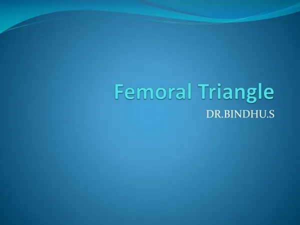
Femoral Triangle. DR.BINDHU.S. Objectives. At the end of the class you will be able to define 1.Femoral triangle 2.Its Boundaries. 3.Contents. 4. Femoral Sheath. 5.Femoral Canal. 6.Applied Anatomy. FEMORAL TRIANGLE.

E N D
Femoral Triangle DR.BINDHU.S
Objectives At the end of the class you will be able to define 1.Femoral triangle 2.Its Boundaries. 3.Contents. 4. Femoral Sheath. 5.Femoral Canal. 6.Applied Anatomy.
FEMORAL TRIANGLE • It is a triangular depression below the inguinal ligament, involves the upper one-third of the front of thigh
Laterally:Medial border of the Sartorius. • Medially:Medial border of the adductor longus. • Base:Inguinal Ligament. • Apex:Point of joining of both the borders.
Boundaries • Roof: • Skin. • Superficial fascia • Superficial inguinal lymph nodes. • Femoral branch of genitofemoral nerve. • Branch of the Illioinguinal nerve. • Superficial branches of the femoral artery • Upper part of the great saphenous vein. • Deep Fascia
FLOOR • Medially by the adductor longus and the pectineus. • Laterally by the iliacus and the psoas major
Contents • Femoral Artery and its branches. • Femoral Vein and its tributaries. • Femoral sheath. • 4 Nerves. • Deep inguinal lymph nodes.
Femoral Artery • Continuation of external iliac artery • 6 branches (3 Superficial and 3 deep): • Superficial External Pudendal. • Superficial Epigastric • Superficial Circumflex iliac. • Profunda femoris. • Medial and lateral circumflex artery. • Deep external pudendal • Muscular branches.
External iliac artery Inguinal ligament Medial circumflex femoral artery Lateral circumflex femoral artery Femoral Artery Profunda femoris artery Descending genicular artery FEMORAL ARTERY
FEMORAL NERVE • Largest br.of lumbar plexus • Root value- L2,L3,L4 • Course- trunk at the iliac fossa. • Enters femoral triangle behind inguinal ligament. • Lateral to the artery. • Outside the sheath. • Between the iliacus and the psoas major tendon.
Branches of femoral nerve • From the trunk- to iliacus, pectineus, vascular • branches. • Anterior division- medial cut.nerve of thigh, intermediate cut.nerve of thigh and muscular br.fr sartorius • Posterior division- saphenous nerve,muscular br.for quadriceps femoris
SAPHENOUS NERVE • Longest cutaneous nerve of the body. • At femoral triangle- Lateral side of femoral artery • Crosses from lateral to medial side in the middle of adductor canal. • In the roof of adductor canal it gives a br. for subsartorial plexus of nerves. • At posteromedial surface of knee, it gives an infrapatellar br. • It accompanies greatsaphenous vein on the medial side of tibia • In the lower third of the leg it divides into two branches. • 1. one follows the medial border of tibia and extend upto the ankle. • 2. 2nd one passes in front of ankle and supplies the skin on the medial side of dorsum of the foot upto first metatarso –phalangeal joint (ball of great toe)
Muscular br. Saphenous nerve
Femoral vein Femoral Vein and its tributaries Femoral vein • Accompanies the artery. • Medial at the base. • Posteriomedial to the artery in the apex. • Tributaries: • Great saphenous vein • Circumflex vein. • Veins corresponding to the branches of the femoral artery.
Femoral Sheath • Encloses upper 3 – 4cms of the femoral vessels. • Extension of two layers of the fascia of the abdomen i.e. fascia transversalis and fascia iliacus. • Devided into 3 compartments: • Lateral / Arterial compartment: • Femoral artery. • Femoral branch of the genitofemoral nerve. • Intermediate / Venous compartment. • Femoral vein. • Medial / Lymphatic compartment: • Femoral Canal.
Femoral Sheath Fascia Transversalis
Femoral canal • Shortest medial compartment of femoral sheath • Base oval shaped & called FEMORAL RING Boundaries: • Laterally- partition between femoral canal and femoral vein. • Posteriorly- Pectineus covered by pectineal fascia • Medially- lacunar ligament • Anteriorly- medial part of inguinal ligament Fascia covering Pectineus Inguinal ligament Lacunar Ligament
Femoral Canal. • 1.5 cm long and wide at the base. • Femoral ring. • Anteriorly: Inguinal ligament. • Posteriorly: Pectineus muscle and fascia. • Medially: Concave margin of the lacunar ligament. • Laterally: Septum seperating it from femoral vein. • Femoral ring closed by the condensation of extraperitoneal connective tissue – Femoral septum. • Contents: • Lymph Node of cloquet or of Rosenmuller, lymphatics and areolar tissue.
Applied Anatomy. • Femoral Hernia.
FEMORAL HERNIA • COVERINGS- • PERITONEUM • FEMORAL SEPTUM • ANT.WALL OF FEMORAL SHEATH • CRIBRIFORM FASCIA • SUPERFICIAL FASCIA • SKIN
Important questions • Boundaries &contents of femoral triangle • Femoral Sheath. • Femoral Canal. • Femoral artery • Femoral nerve • Femoral vein • Saphenous nerve • Femoral hernia