Tissues: Living Communities
1.01k likes | 1.03k Vues
Learn about the functions of epithelial tissues, differentiate between types of cellular junctions, describe the structure of the basement membrane, and classify different epithelial tissues and glands.

Tissues: Living Communities
E N D
Presentation Transcript
Learning Objectives Describe the functions of epithelial tissues. Differentiate between the three major types of cellular junctions found between epithelial cells. Describe the structure of the basement membrane. List and describe the characteristics used to classify different epithelial tissues. List and describe the characteristics used to classify different glands. List and describe the components that make up connective tissues. Differentiate between areolar, adipose, and reticular connective tissues. Differentiate between dense regular, dense irregular, and elastic connective tissues. Differentiate between hyaline cartilage, elastic cartilage, and fibrocartilage. List and describe the components of bone.
Types of Tissues • Tissues are classified into the following four primary types: 1. Epithelial tissue (covers and lines) 2. Connective tissue (provides support) 3. Muscle tissue (enables movement) 4. Nervous tissue (controls work)
Epithelial TissuesFunctions: Sheets of cells that cover and line other tissues Protect underlying tissues and may act to filter biochemical substances May absorb, secrete, or excrete biochemical substances May play a role in the reception of sensory input
Characteristics of Epithelia • Each epithelial cell has an apical surface and a basal surface • Apical surface faces the lumen or outside of the organ • Basal surface faces the basal lamina and blood vessels • Lateral surfaces are connected to neighboring cells by junctional complexes. • Epithelial cells are avascular. • Most epithelial cells are innervated.
Cellular Attachments Three major types of cellular junctions 1. Tight junctions 2. Desmosomes 3. Gap junctions
Tight Junctions • Formed by the fusion of the outermost layers of the plasma membranes of adjoining cells • Found in tissues in which there can be no leaks (urinary bladder, digestive tract)
Desmosomes • Mechanical coupling formed by filaments that interlock with one another (kind of like velcro) • Tonofilaments extend from the plaque into the cytoplasm. • Found in tissues that undergo repeated episodes of tension and stretching (skin, heart, uterus) • Hemidesmosomes link epithelial cells to the basement membrane.
Gap Junctions • Tubular channel proteins (called connexons) that extend from the cytoplasm of one cell to the cytoplasm of another • Allow exchange and passage of ions and nutrients • Found in intestinal epithelial cells, the heart, and smooth muscle tissue
Basement Membrane • Meshwork of fibers that cements the epithelial cell to the underlying connective tissue • Also called basal lamina • Varies in thickness • Helps prevent the cell from being torn off by intraluminal pressures • Acts as a partial barrier between the epithelial cell and the underlying connective tissue
Surface Specialization • Surfaces of epithelial cells vary depending on where they are located and what role they play in the function of the tissue • Smooth (Blood vessels) • Microvilli (called the brush border)-increases surface area for absorption • Cilia • Keratin (water proof).
Classification of Epithelial Tissue Number of layers of cells: Simple or stratified Shape of the cells: Squamous, cuboidal, and columnar Presence of surface specializations: Cilia, keratin, etc.
Simple Squamous Epithelium Fragile and thin Found lining surfaces involved in the passage of either gas or liquid Flat and smooth
Simple Cuboidal Epithelium Single layer of cube-shaped cells Round, dark-staining nuclei aligned in a single row Occurs in areas of the body where secretion and absorption take place
Simple Columnar Epithelium Elongated and closely packed together Nuclei aligned in a row at the base of the cell near the basement membrane Found in many excretory ducts as well as in the digestive tract
Stratified Squamous Epithelium Multilayered Occur in areas of the body subject to mechanical and chemical stresses Protect underlying tissues
Stratified Cuboidal Epithelium Usually two layers of cuboidal cells Found primarily along large excretory ducts Protects underlying tissues
Stratified Columnar Epithelium Found only in select parts of the respiratory, digestive,reproductive systems and along some excretory ducts Function in secretion and protection
Pseudostratified Columnar Epithelium Cell nuclei are found at different levels across the length of the tissue Some cells do not reach the luminal surface Found in respiratory tract and in portions of the male reproductive tract
Transitional Epithelium Stratified epithelium with a basal layer of cuboidal or columnar cells and a superficial layer of cuboidal or squamous cells Found in areas of the body required to expand and contract as part of their normal function
Glandular Epithelium • Groups of cells that manufacture and discharge a secretion • Classification of glands • Presence or absence of ducts • Number of cells that compose them • Shape of the secreting ducts • Complexity of the glandular structure • Type of secretion they produce • Manner in which the secretion is stored and discharged
Endocrine Glands Glands that do not have ducts or tubules / secretions are distributed throughout the body Produce and secrete hormones into the bloodstream or the lymphatic system Part of a complex, biochemical network known as the endocrine system which includes pituitary gland, adrenal gland, parathyroid gland, pancreas, thyroid gland, testes, ovaries,
The endocrine glands are made up of simple epithelial cells… Pituitary gland
Goblet Cell Single cell exocrine gland Ductless and composed of modified columnar epithelial cell Found among columnar cells of the respiratory and digestive tracts and the conjunctiva of the eye Secretes mucin Provides lubrication and “entrapment”
Exocrine Glands Discharge secretions via ducts directly into local areas (except for goblet cell-no duct and endocrine glands which have no ducts) Unicellular (goblet cell) or multicellular
Multicellular Exocrine Glands Composed of a secretory unit and a duct Secretory unit is usually surrounded by connective tissue rich in blood vessels and nerve fibers
Classification of Exocrine Glands • Ducts: • Simple: main duct is unbranched • Compound: main duct is branched • Shape of secretory portions • Tubular: secretory cells form a long channel of even width • Alveolar or acinar: secretory unit forms a rounded sac • Tubuloalveolar, or tubuloacinar: secretory units possess both tubular and alveolar qualities
Classification of Exocrine Glands Merocrine glands package their secretions and release them via exocytosis as they are manufactured. Apocrine glands store their secretions and then release the top part of the cell into the duct system Holocrine glands store their secretions and then release the entire contents of the cell
Classification of Exocrine Glands Type of secretion produced • Serous secretions • Watery • Contain a high concentration of enzymes • Mucous secretions • Thick, viscous • Composed of glycoproteins. • Protects apical layer (intestines), traps microorganism so cilia can move them out (respiratory) • Mixed exocrine glands contain both mucous and serous components (digestive and respiratory.
Connective Tissue Functions-different types of connective tissue: Form metabolic and structural connections between other tissues…support Forms a protective sheath around organs and helps insulate the body…cushion Acts as a reserve for energy…storage Provides the frame that supports the body…support Composes the medium that transports substances from one region of the body to another…transport Plays a role in the healing process and in the control of invading microorganisms…repair and defense
Connective Tissue Components • Extracellular matrix • Extracellular fibers • Ground substance • Cells
Connective Tissue Components Ground substance Medium through which cells exchange nutrients and waste with the bloodstream Amorphous, homogeneous material Ranges in texture from a liquid or gel to a calcified solid Made up of glycosaminoglycans, proteoglycans, glycoproteins Serves as an effective obstacle for invading microorganisms
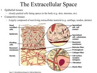
Connective Tissue Components Extracellular fibers • Collagenous fibers • Strong, thick strands of collagen • Organized into bundles of long, parallel fibrils composed of bundled microfibrils • Variable density and arrangement of fibers • Found in tendons and ligaments
Connective Tissue Components Extracellular fibers • Reticular fibers • Thin, delicate, branched networks of collagen • Provide support for highly cellular organs (endocrine glands, lymph nodes, spleen, bone marrow, and liver) • Also found around blood vessels, nerves, muscle fibers, and capillaries
Connective Tissue Components Extracellular fibers • Elastic fibers • Branched networks composed primarily of the protein elastin • Composed of coiled bundles of microfibrils • Occur in tissues commonly subjected to stretching (vocal cords, lungs, skin, and walls of blood vessels)
Connective Tissue Components Cell Types • Fixed Cells: involved in production and maintenance of the matrix • Fibroblasts, chondroblasts, osteoblasts, adipocytes, reticular cells FIBROBLASTS
Fibroblasts A fibroblast is a cell actively producing collagen fibers and matrix meterial. They are protein secreting cells. In intermuscular CT- Forms scar tissue in the muscle where there is injury. Scar tissue- collagen fibers…fibrosis Wound repair Characteristics: Thin and flattened “arms” to rest on matrix structure-lie on top Not a lot of cytoplasm so they look like spindle shaped nuclei lying along the fibers.
Transient Cells • Transient/ Wandering Cells: involved in the repair and protection of tissues • Leukocytes, mast cells, macrophages • Move in and out of connective tissue - Go where needed as needed • Leukocytes-”wander” into the bloodstram from connective tissue • Mast cells-histamine and heparin…tend to be near blood vessels • Macrophages-inflammation • Resident macrophages in the lymphatic organs, lungs, liver, spleen, lamina propria of digestive tract
Types of Connective Tissue Blood Lymphatic Plasma Bone Cartilage CT Proper Loose CT Dense CT Areolar Adipose Reticular Dense regular Dense irregular Elastic
Connective Tissue Proper • Loose connective tissue • Areolar • Adipose • Reticular • Dense connective tissue • Dense regular • Dense irregular • Elastic
Areolar Connective Tissue • Loose connective tissue • Fibers and cells suspended in a thick, translucent ground substance • More cells vs not a lot of fibers • Predominant cell is the fibroblast (flat) • Manufactures the elastic, reticular, and collagenous fibers • Surrounds every organ; forms the SQ layer that connects skin to muscle; envelopes blood vessels, nerves, and lymph nodes; present in all mucous membranes
Areolar Connective Tissue Description Loose array of fibers Includes all three types of fibers Many cells Location Under epithelial basement membranes Between glands muscles nerves Surrounding capillaries Surrounding organs Under skin Function Provides nutrients to tissues Supports “packing material”
Adipose Connective Tissue Areolar tissue in which adipocytes predominate Loose connective tissue with little to no matrix/ very cellular Two types- white adipose/ brown The cells are filled with lipids -differences
White vs Brown adipose • Amount of lipid fluctuates, cell # stays the same • Nuclei get “pushed” to side • Food animals (and other animals) store pesticides and drugs in the adipose…withdrawl mandated by law for food animals • Very vascular • Lots of mitochondria • Brown color • Thermogenic-generates heat • Rodents that hibernate
Adipose Connective Tissue Function Protects organs and other tissues Thermoinsulator Energy storage Description Very little extracellular material Adipocytes filled with lipids Nuclei and organelles are pushed to side “Chicken wire” appearance Location Beneath the skin Spaces between muscles Behind eyeballs Surface of the heart Around the kidneys and heart Surrounding joints In bone marrow In the omentum of the abdomen Around the colon
Reticular Connective Tissue Loose connective tissue Network of thin reticular fibers…structural support not strength Contains loosely arranged fibers and many fibroblasts suspended in a supportive ground substance Forms the stroma(framework)(spleen, lymphatic organs)
Reticular Connective Tissue Description Loosely arranged fibers Reticular fibers only fibroblasts Location Spleen Lymph nodes Bone marrow liver Function Provides framework (stroma)