Diabetic Foot
700 likes | 1.64k Vues
Diabetic Foot. Diabetic Foot. Any foot pathology that results directly from diabetes or its long term complications ( Boulton 2002)

Diabetic Foot
E N D
Presentation Transcript
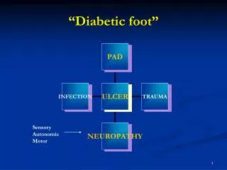
Diabetic Foot • Any foot pathology that results directly from diabetes or its long term complications ( Boulton 2002) • The foot of a diabetic patient that has the potential risk of pathologic consequences including infection, ulceration and or destruction of deep tissues associated with neurologic abnormalities, various degrees of peripheral vascular disease and/or metabolic complications of diabetes in the lower limb
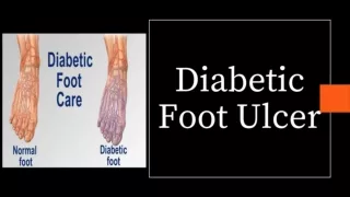
Diabetic Foot • Diabetic foot ulcer • Diabetic foot infections • Charcot Joints
Epidemiology • DM is the largest cause of neuropathy • 50% patients don’t know that they have diabetes • Foot ulcerations is most common cause of hospital admissions for Diabetics • Expensive to treat, may lead to amputation and need for chronic institutionalized care
Pathophysiology • Combination of factors • Neuropathy • Peripheral arterial disease • Abnormal foot biomechanics • Delayed wound healing
Diabetic Neuropathy • Microvascular complication • Occlusion of vasa nervosum • Can be • Sensory / motor/ autonomic • Mono / poly / radiculopathy • Most commonly distal symmetric sensory neuropathy
Neuropathy • Sensory Neuropathy • Loss of touch and temperature • Minor trauma goes unnotices • Disorders of proprioception • Abnormal weight bearing • Callus formation, ulceration • Motor and sensory neuropathy • Abnormal foot biomechanics • Structural changes
Neuropathy • Autonomic neuropathy • Anhidrosis in lower limbs • Drying of feet • Fissure formation
Altered biomechanics • Abnormal weight bearing • Fixed foot deformities • Hammer toe • Claw toe • Prominent metatarsal heads • Charcot’s joints
Hammer Toes Claw Toes
Other factors • Impaired wound healing • Does not allow resolution of fissures and minor injuries • Increased chances of infection
Peripheral arterial disease • 30 times more prevalent in diabetics • Diabetics get arteriosclerosis obliterans or “lead pipe arteries” • Calcification of the media • Often increased blood flow with lack of elastic properties of the arterioles • Not considered to be a primary cause of foot ulcers
Causal Pathways for Foot Ulcers % Causal Pathways Neuropathy: 78% Minor trauma: 79% Deformity: 63% Behavioral ? Neuropathy Deformity Minor Trauma - Mechanical (shoes) - Thermal - Chemical Poor self-foot care ULCER
Risk Factors for Diabetic Foot • Male Sex • DM > 10 years duration • Peripheral neuropathy • Abnormal foot structure • Peripheral arterial disease • Smoking • H/O previous ulceration / amputation • Poor glycemic control
Examination • Neurological examination • Vibration perception • Light pressure • Light touch • Two point discrimination • Pain • Temperature perception • Deep tendon reflexes • Clonus • Babinski test • Romberg test • Vascular Examination • Palpation of pulses • Skin/limb colour changes • Presence of edema • Temperature gradient • Skin changes • Atrothy • Abnormal wrinkling • Absence of hair • Onychodystrophy • Venous filling time
Examination • Dermatological • Skin appearance • Calluses • Fissures • Nail appearance • Hair growth • Ulceration/infection/ gangrene • Interdigital lesions • Tineapedis • Markers of diabetes • Musculoskeletal • Biomechanical abnormalities • Structural deformities • Prior amputation • Restricted joint mobility • Tendo Achilles contractures • Gait evaluation • Muscle group strength testing • Plantar pressure assessment
Investigations • Blood investigation • FBS, PPBS • HbA1C • Complete blood counts • ESR • RFT • Urinalysis • Wound / blood culture
Imaging • Plain X-rays • Osteomyelitis • Fractures • Dislocations • Osteolysis • Structural foot abnormalities • Arterial calcification • Tissue gas
Vascular evaluation • Non invasive evaluation • Doppler segmental pressure and waveform analysis • Ankle brachial pressure index • Toe blood pressure • Transcutaneous CO2 • Laser doppler velocimetry
Interpretation of ABI InterpretationABI Normal 0.90-1.30 Mild obstruction 0.70-0.89 Moderate obstruction 0.40-0.69 Severe obstruction <0.40 Poorly compressible >1.30 2° to medial calcification *Poor ulcer healing with ABI < 0.50 **Further vascular evaluation needed
Vascular evaluation • Invasive evaluation • Arteriography • MR angiography • CT angiography
Wagner’s Classification 0 – Intact skin (impending ulcer) 1 – superficial 2 – deep to tendon or ligament 3 - deep abscess, osteomyelitis 4 – gangrene of toes or forefoot 5 – gangrene of entire foot
Treatment • Prevention • Identification of high risk patients • Patient education • Careful selection of foot wear • Daily inspection of feet • Daily foot hygiene • Keep foot clean, moist • Avoidance of self treatment of foot abnormalities and high risk behavior ( walking barefoot) • Prompt consultation with health care provider • Orthotic shoes and devices • Callus management • Nail care
Identifying at risk patient • History: • Prior amputation or foot ulcer • Peripheral artery disease (PAD) • Exam: • Insensate • Foot deformities • Absent pulses • Prolonged venous filling time • Reduced ABI • Pre-ulcerative cutaneouspathology
Treatment • Attention to other risk factors • Smoking • Hypertension • Dyslipidemia • Glycemic control
Treatment • Plantar surface of the foot is the most common site • Ulcer may be • Primarily neuropathic • a/w surrounding cellulitis/ ostemyelitis • Cellulitis without ulceration may occur
Treatment • Offloading • Debridement • Wound dressing • Antibiotics • Revascularisation • Amputation
Treatment • Wagner 0-2 • Total contact cast • Distributes pressure and allows patients to continue ambulation • Principles of application • Changes, Padding, removal • Antibiotics if infected
Treatment • Wagner 0-2 • Surgical if deformity present that will reulcerate • Correct deformity • exostectomy
Treatment • Wagner 3 • Excision of infected bone • Wound allowed to granulate • Grafting (skin or bone) not generally effective
Treatment • Wagner 4-5 • Amputation • ? level
Indications for Amputation • Uncontrollable infection or sepsis • Inability to obtain a plantar grade, dry foot that can tolerate weight bearing • Non-ambulatory patient • Decision not always straightforward
Treatment • After ulcer healed • Orthopedic shoes with accommodative (custom made insert) • Education to prevent recurrence
Wound Care products • Dressings • Gauze pads • Transparent films • Hydrogels • Foam • Hydrocolloid • Alginate • Collagen dressing • Antimicrobial dressings